Pseudoophiobolus rosae Phookamsak, Wanas., Phukhamsakda, Camporesi & K.D. Hyde, sp. nov. Index Fungorum number: IF553928
Etymology: The specifific epithet ‘‘rosae’’ refers to the host Rosa
Holotypus: MFLU 15-1014
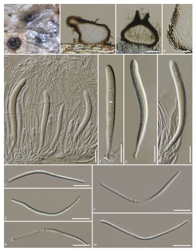
Saprobic on Rosa canina L. Sexual morph: Ascomata 280–350 µm high (including papilla), 210–340 µm diam., dark brown to black, scattered, solitary, semi-immersed in the host epidermis, becoming superficial when the epidermis was removed, ampulliform, cupulate when dried, uniloculate, glabrous, with dark brown, septate mycelium at the base, adhering to the host, ostiolate, papillate. Papilla 100–140 µm high, 90–110 µm diam., mammiform, apex rounded, short papillate, composed of several layers of dark pseudoparenchymatous cells, arranged in a textura angularis, ostiole central, with pore-like opening and hyaline periphyses. Peridium 20–45 µm wide, slightlyk, composed of several layers of hyaline to dark brown, thin-walled, pseudoparenchymatous cells, inner layers comprising 2–3 layers of hyaline cells, arranged in textura angularis, outer layers comprising 3–5 layers of dark brown to black, thick-walled cells, arranged in textura angularis to textura globulosa. Hamathecium composed of dense, 1.5–3 µm wide, cellular pseudoparaphyses, indistinctly septate, not constricted at the septa, anastomosing at the apex, embedded in a hyaline gelatinous matrix. Asci (85–)90–130(–142) x (8–)9–12(–13) µm (x = 112.6 x 11.2 µm, n = 25), 8-spored, bitunicate, fifissitunicate, cylindric-clavate, short pedicellate, apically rounded, with well-developed ocular chamber. Ascospores (75–)80–100(–108) x 2–3.5 µm (x = 92.5 x 3 µm, n = 30), fasciculate, scolecosporous, pale yellowish to yellowish, subcylindric-clavate to fifiliform, with rounded ends, tapered towards the lower cells, swollen at the 8th, or 10th cell, 15(–22)-septate, not constricted at the septa, smooth-walled. Asexual morph: Undetermined.
Culture characteristics: Colonies on PDA reaching 52–55 mm diam. after 4 weeks at 25–30 C; medium dense, circular, flflattened, surface smooth, edge entire, velvety to cottony, with small turfs at the centre, colony from above cream, yellowish sectors at the centre, colony from below, cream at the margin, pale yellowish to pale brown at the centre, not producing pigmentation in agar.
Material examined: ITALY, Province of Arezzo [AR], Bagno di Cetica, dead spines of Rosa canina L. (Rosaceae), 13 October 2014, E. Camporesi, IT 2171 (MFLU 15-1014, holotype) ex-type living culture, MFLUCC 17-1786 = KUMCC 17-0170
Notes: Pseudoophiobolus rosae is similar to P. achilleae and P. subhyalinisporus in having subhyaline to pale yellowish ascospores, but can be distinguished from these species in having more septa in the ascospores, ascospores narrowed towards the lower end cells and swollen at the 8th or 10th cell (Table 3). Ascospore septation of P. rosae is indistinct, whereas it is distinct in P. achilleae and P. subhyalinisporus (Figs. 8, 13). Ophiobolus fruticum has been reported on Rosa sp. from Ukraine (Farr and Rossman 2017). However, the species differs from Pseudoophiobolus rosae in having ascospores with more septa and with three swollen cells. Multigene phylogenetic analyses show that Pseudoophiobolus rosae clusters with P. achilleae, P. erythrosporus, P. galii, and P. urticicola.
Hosts: Rosa canina L. (Rosaceae)
Distribution: Italy
FIG Pseudoophiobolus rosae (MFLU 15-1014, holotype). a Appearance of ascomata on the host surface. b, c Section through ascoma. d Section through peridium. e Asci with pseudoparaphyses. f–h Asci. i–m Ascospores. Scale bars: b, c = 100 µm, d–m = 20 µm