Pseudocamarosporium pinicola Wijayaw., Camporesi & K.D. Hyde, sp. nov., Index Fungorum number: IF551302
Etymology: Named after the generic name of the host
Endophytic or saprobic on dead branch of Pinus nigra. Sexual morph: Undetermined. Asexual morph: Conidiomata 1000–1300 μm diam., 800–1100 μm high, pycnidial, immersed, globose to subglobose, unilocular, solitary, black, with a papillate, centrally located ostiole. Pycnidial wall multi-layered, with a thick outer layer, composed of dark brown cells of textura angularis, with a thick, hyaline inner layer with cells of textura angularis. Conidiophores reduced to conidiogenous cells. Conidiogenous cells 19–21 × 16–20 μm, cylindrical to ampulliform, holoblastic, phialidic, discrete, determinate, hyaline, smooth, formed from the inner layer of the pycnidial wall. Conidia 35–46 × 19–24 μm (mean = 40.38 × 22.02 μm, n = 20), ellipsoid to oblong, muriform, with 3 transverse and 2 longitudinal septa, continuous, straight, occasionally slightly curved, golden to dark brown, smooth-walled.
Culture characteristics: Colonies on PDA slow-growing, reaching 2 cm diam. after 1 week at 18 °C, thin mycelium, circular, rough margin white at first, white from the top, pale brown from reverse after 1 week, flat or effused on the surface. Hyphae septate branched, hyaline, thin.
Material examined: ITALY, Province of Forlì-Cesena [FC], Corniolo – Santa Sofia on dead branch of Pinus nigra (Pinaceae), 7 December 2013, Erio Camporesi, IT 1564 (MFLU 15–0735, holotype) (HKAS 88743); living cultures MFLUCC 14–0457, GUCC 60.
Notes: – Pseudocamarosporium brabeji Marinc. et al. (14–18.5 × 7–9 μm fide Marincowitz et al. 2008). Camarosporium pini (Westend.) Sacc. (18–20 × 9–10 μm fide Barna et al. 2010) and Camarosporium propinquum (Sacc.) Sacc. have been reported previously from Pinus spp. (both species on Pinus halepensis fide Farr and Rossman 2016). Both these species are morphologically distinct from our collection. In molecular data analyses, our new strain resides in Pseudocamarosporium sensu stricto, but is phylogenetically distinct from P. propinquum. Therefore, our collection is introduced as a new species.
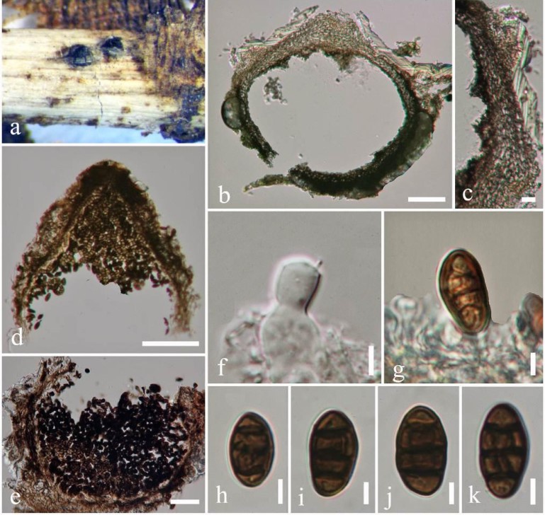
Fig. 3 Pseudocamarosporium pinicola (holotype) a Conidiomata on dead stem of Pinus nigra. b, d, e Vertical sections of pycnidia. c Conidioma wall. f-g Conidiogenous cells and conidiogenesis. h-k Conidia. Scale bars: b = 200 μm, c = 40 μm, d–e = 100 μm, f–k = 10 μm.