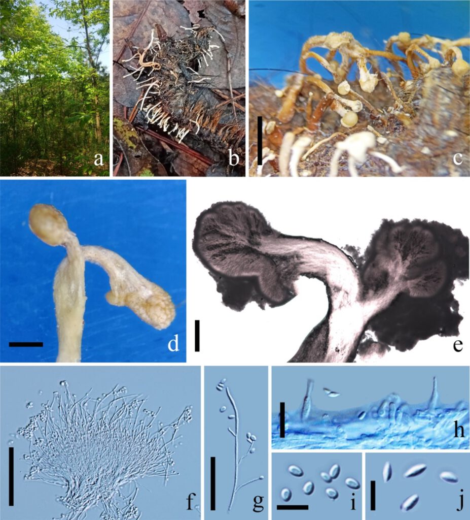
Pleurocordyceps sinensis (Q.T. Chen, S.R. Xiao & Z.Y. Sh) Y.J. Yao, Y.H. Wang, S. Ban, W.J. Wang, Yi Li, Ke Wang & P.M. Kirk, in Wang, Ban, Wang, Li, Wang, Kirk, Bushley, Dong, Hawksworth & Yao, Journal of Systematics and Evolution: 10.1111/jse.12705, 11 (2021). Fig. 17.
MycoBank number: MB 570676; Index Fungorum number: IF 570676; Facesoffungi number: FoF 10739;
Parasitic on larvae of Lepidoptera, on leaf litter. Sexual morph: Undetermined. Asexual morph: Hyphomycetous. Synnemata 0.2–1 cm long, 0.2–4 mm wide, cylindrical, clavate, capitate, stipitate, solitary to crowded, simple or branched, brown to yellow to yellowish, with or without fertile head. Stipe 0.2–1 cm long, 1–2 mm wide, cylindrical, brown to yellow, with or without fertile head. Fertile head 0.2–4 mm wide, globose to subglobose, yellowish, with conidia masses covered. Phialides two types, both types observed on the same synnema. α-phialides 10–24 × 0.9–1.2 μm ( = 17 × 1.1 µm, n = 90), gathered on the topical of synnema, arranged in a parallel palisade-like layer around the top of fertile head, hyaline, usually branched into 2–6 phialides, narrow slender lanceolate; β-phialides 10–16 × 1.1–1.8 μm ( = 13 × 1.5 µm, n = 90), solitary, scattered along the stipe, lanceolate, ovate at the base, 6–12 μm long, tapering from into a long neck, 2–4 μm long. Conidia one-celled, hyaline, smooth, two types. α-conidia 1.7–2.4 μm ( = 2.1 µm, n = 90), spherical, forming slime conidial masses on the fertile head; β-conidia 3–5 × 1.4–2.1 μm ( = 4 × 1.7 µm, n = 90), fusiform, produced along stipe of the synnema.
Colonies on PDA medium, growing slowly, attaining 5 cm in 20 days at 25°C, white, reverse yellow. Synnemata emerging after 20 days, 0.3–1 cm long, solitary, white, scattered on the surface of colony, branched or unbranched, without fertile head. Conidial masses generating from the apex of the synnema. Conidiophores undetermined, not clear. Phialides existing in two types: α-and β-phialides. α-phialides 10.4–18.3 × 0.8–1.8 μm ( = 14.4 × 1.3 µm, n = 90) hyaline, narrow slender, smooth. β-phialides 22.9–64.2 × 1.0–1.5 μm ( = 43.6 × 1.3 µm, n = 90) solitary, growing from hyphae, narrow slender, catenate, smooth. α-conidia 1.8–2.2 × 1.4–1.9 μm ( = 2 × 1.7 µm, n = 90) globose to subglobose, occurring in the conidial mass on the agar or on the final portion of synnemata, one-celled, smooth-walled, yellow slimy in mass. β-conidia 3.2–4.1 × 1.4–1.8 μm ( = 3.6 × 1.6 µm, n = 90) fusiform, and produced on the surface mycelium of colony or on the top of the synnemata, one-celled, smooth-walled, hyaline, usually in chains on a phialide.
Material examined: China, Guizhou Province, Zunyi City, Wuchuan County. Parasitic on larvae of Lepidoptera, on the leaf litter, 23 October 2019, Yu Yang (New host: GZU 22-ZNR0219102301).
Notes: The strains are with the same group with Pleurocordyceps sinensis and form an Pleurocordyceps sinensis clade (Fig. 16). Moreover, the morphology characters are the same as Pleurocordyceps sinensis. These strains are identified as Pleurocordyceps sinensis based on morphology and phylogenetic analyses. However, the host of those strains is different from the holotype of Pleurocordyceps sinensis, which parasitizes Ophiocordyceps sinensis. Larvae of Lepidoptera (Fig. 17: GZU 22-ZNR0219102301, Fig. 18: MFLU 21-0269), Ophiocordyceps crinalis (Fig. 20: GZU-520326MF865c) and Ophiocordyceps barnesii (Fig. 21: FC20042304) are the new hosts of Pleurocordyceps sinensis. These specimens were collected from Guizhou and Yunnan, China. We introduced the new host for Pleurocordyceps sinensis, and this study shows the geographic distribution of Pleurocordyceps sinensis, illustrating its broad host range. However, these strains share the same clade with Polycephalomyces ramusus and Polycephalomyces tomentosus, with similar morphology. The representative strains of Polycephalomyces ramusus and Polycephalomyces tomentosus lack morphological details and requirie further morphology information (Wang et al. 2021). Thus, these strains were identified as Pleurocordyceps sinensis.

Fig. 17. Pleurocordyceps sinensis (New host ZNR0219102301) a Habitat of Perennicordyceps sinensis. b Overview of host. c-e Synnemata from infected Polycephalomyces sinensis. f Conidiophores. g α-phialides. h β-phialides. i α-conidia. j β- conidia. Scale Bars: c = 2000 µm, d, e = 1000 µm, f = 50 µm, g = 20 µm, h = 10 µm, i, j = 5 µm.
