Phyllosticta doitungensis Bhunjun & K.D. Hyde, sp. nov.
MycoBank number: MB 559262; Index Fungorum number: IF 559262; Facesoffungi number: FoF 10581; Fig. 1
Etymology: The epithet refers to the type locality.
Holotype: MFLU 21-0175
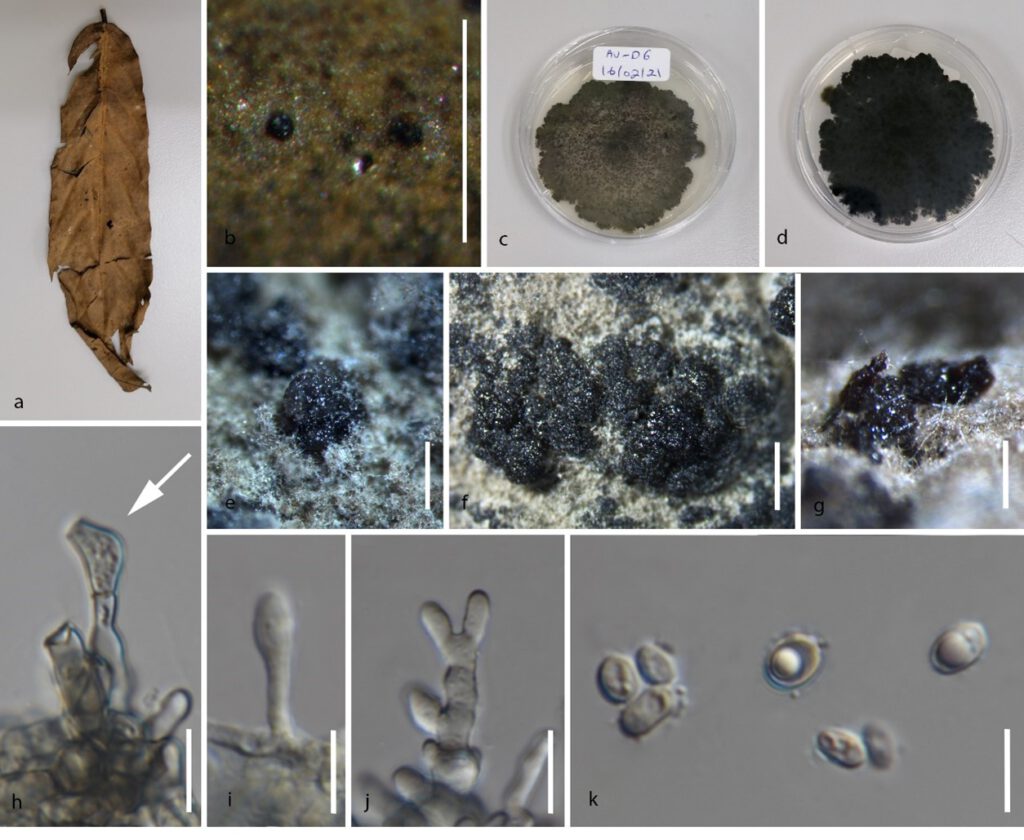
Saprobic on decaying leaf of Dasymaschalon obtusipetalum Wang, Chal., Saund. Sexual morph: not observed. Asexual morph on host and culture: Conidiomata (on host) pycnidial up to 100 μm diam, solitary, black, semi-immersed, globose. Conidiomata (on PDA) up to 600 μm diam, solitary or arranged in clusters of up to 3, black, globose to ampulliform, base ovoid, including colorless to opaque conidial masses. Pycnidial wall consisting of several layers, up to 30 μm thick, outer layer of dark brown textura angularis, inner wall of 1–2 layers of hyaline textura angularis cells. Ostiole single, central, up to 20 μm diam. Conidiophores reduced to conidiogenous cells, subcylindrical to ampulliform. Conidiogenous cells 6−8 × 2–4 μm, terminal, subcylindrical, hyaline, smooth, proliferating several times percurrently near apex. Conidia 10–13 × 7–9 μm (x̅ = 11 × 8 μm, n = 20), solitary, hyaline, aseptate, thin and smooth walled, with a single large central guttulate, broadly ellipsoid to ovoid, tapering towards a bluntly rounded apex, forming appressoria within 2 days, apical mucoid appendage not observed.
Culture characteristics: On PDA, colonies flat, fast growing reaching 4 cm after 14 days, initially white with abundant mycelium, lobate margins, gradually becoming grey after 2–3 days with white hyphae around the margin, eventually turning black; reverse black after 14 days.
Habitat: on leaf of Dasymaschalon obtusipetalum.
Distribution: Thailand.
Material examined: THAILAND, Chiang Rai Province, Doi Tung, on leaf of Dasymaschalon obtusipetalum, 31 Jul 2020, CS Bhunjun, AV–D6 (MFLU 21-0175, holotype), ex-type living culture, MFLUCC 21-0074.
GenBank numbers: ITS: XXXX, LSU: XXXX, tef: XXXX, rpb2: XXXX.

Fig. 1 Phyllosticta doitungensis (MFLU 21-0175, holotype). a Host. b Appearance of conidiomata on the host leaf surface. c, d Colony on PDA 14 days after inoculation. e–g Formation of pycnidia on PDA. h Developing appressoria. i–j Conidiogenous cells giving rise to conidia. k Conidia. Scale bars: b, e–g = 500 μm, h–k = 10 μm.


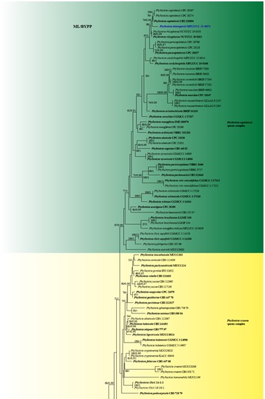
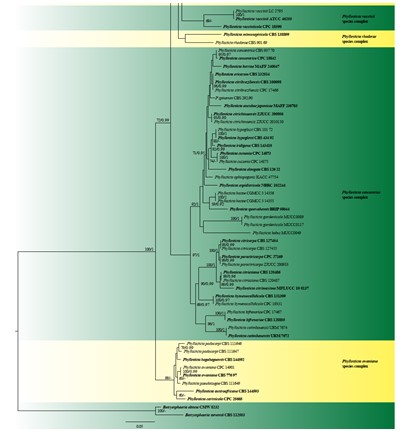
Fig. 2 The best scoring RAxML tree based on a combined ITS, LSU, tef, act, gapdh and rpb2 dataset. Related sequences were retrieved from GenBank and Norphanphoun et al. (2020). One hundred and twenty-six strains were included in the analysis of the combined loci which comprised 3127 characters after alignment. The tree is rooted with Botryosphaeria obtusa (CMW 8232) and B. stevensii (CBS 112553). The best scoring RAxML tree had a final likelihood value of -23828.412687. Estimated base frequencies were: A = 0.213306, C = 0.290222, G = 0.278595, T = 0. 0.217877; substitution rates AC = 1.098874, AG = 3.337079, AT = 1.201287, CG = 1.057992, CT = 6.887752, GT = 1.000000, gamma distribution shape parameter α = 0.279545. Maximum likelihood (ML) bootstrap values higher than 50% and Bayesian posterior probabilities (BYPP) greater than 0.90 are given at the nodes. Hyphens (-) represent support values less than 50% BS/0.90 BYPP. The ex-type strains and reference strains are in bold. The newly generated sequence is in blue.
