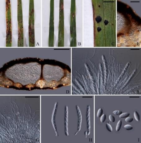
Phyllachora siamensis Maharachch., K.D. Hyde & Cheew, sp. nov.
MycoBank number: MB 840846; Index Fungorum number: IF 840846; Facesoffungi number: FoF 10632; Fig. **
Etymology: Refers to the country Thailand, where the specimen was collected.
Holotype: CMU TAR08
Associated with the blackened spots on living leaves of Chloris barbata SW. and Eleusine indica L. Sexual morph on host: Tar spots 0.2–0.5 × 0.2–1 mm, on upper and lower leaves surface, as black punctiform spots, ovoid, shiny, with raised glossy black clypeus covering the ascomata. Stromata uni-multiloculate, 340–(520–610)–700 µm wide, 162–(200–250)–290 µm high, ovoid to globose, on the upper leaves surface, the stroma immersed with the upper wall of the ascoma, black thick-walled completely occluded by melanin. Ascomata perithecial, 155–(195–232) wide, 105–(157–182)–210 µm high, flask- to bowl-shaped, solitary or forming groups of 1–2, globose, the ostiole inconspicuous, not or only weakly papillate. Peridium composed of thin, dark brown layers. Paraphyses longer than mature asci, filiform, branched, septate, not constricted at the septate. Asci 62–(80–95)–105 × 7–10 µm, 8–spored, unitunicate, clavate to cylindrical. Ascospores10–13 × 5–6 µm, overlapping un-biseriate, unicellular to ovate, smooth, hyaline, surrounded by mucilaginous sheath. Asexual morph: Not observed.
Habitat: on living leaves of Eleusine indica and Chloris barbata.
Distribution: Thailand.
Material examined: Thailand, Chiang Mai, on living leaves of Eleusine indica (Poaceae), 2.8.2016, N. Tamakaew (CMU TAR08, holotype); ibid., Royal Project Foundation of Nonghoy, 18º93ʹ0ʺN, 98º82ʹ47ʺE, on living leaves of Chloris barbata (Poaceae), N. Tamakaew (CMU TAR12).
GenBank numbers: CMU TAR08 (ITS: MZ749653, LSU: MZ749659, SSU: MZ749661), CMU TAR12 (ITS: MZ749654, LSU: MZ749660, SSU: MZ749662)
Notes: Phyllachora siamensis was isolated from living leaves of Eleusine indica and Chloris barbata in Thailand and forms a sister clade to Phyllachora chloridis (MFLU 15-0173 and MFLU 16-2980) (100% ML, Fig. 12) which were isolated from Chloris sp. in Thailand (Dayarathne et al. 2017). Phyllachora siamensis can be distinguished from P. chloridis in having larger ascomata (195–232 × 157–182 vs 80–90 × 90–100 µm), larger asci (80–95 × 7–10 vs 50–72 × 6–8) and broader ascospores (10–13 × 5–6 µm vs 8–12 × 3.5–4.8 µm). Furthermore, a guttule and a mucilaginous sheath were observed in the ascospores of P. chloridis, which were not observed in P. siamensis. Dayarathne et al. (2017) proposed Neophyllachora to accommodate Phyllachora cerradensis, P. myrciae, P. myrciariae, P. subcircinans and P. trucantispora which formed a strongly supported monophyletic clade separate from other Phyllachora species. However, in our phylogenetic analysis, we find evidence for non-monophyly of Phyllachora and therefore, the generic status of Neophyllachora should be evaluated.

Fig. X Phyllachora siamensis (CMU TAR08, holotype). A, B Tar spots on upper surface. C Close up of tar spots on the leaf. D, E Vertical section through tar spot illustrating central ostiole, peridium and position in host tissues. F Asci and paraphyses. G Paraphyses. H Asci. I Ascospores. Scale bars: C = 5 mm, D = 100 µm, E = 30 µm, F = 20 µm, G = 5 µm, H = 20 µm, I = 10 µm.
