Phaeosphaeriopsis dracaenicola Phookamsak & K.D. Hyde, sp. nov., Index Fungorum number: IF550737
Etymology: referring to the host, of which the fungus was found.
Holotypus: MFLU 11-0193
Pathogenic causing necrotic leaf spot on Dracaena lourieri; lesions 3–6 cm, infecting mostly on leaf margin, oval-shaped, pale brown to yellowish-brown, separated from healthy tissue by a reddish-brown to orange-brown margin. Sexual state: Ascomata 88.5–170 µm high, 140–200 µm diam., solitary to gregarious, immersed, visible as slightly raised, small black spots on host surface, subglobose, brown to dark brown, ostiole central. Peridium 5–17.5 µm wide, thin-walled, of equal thickness, composed of 3–4 layers of dark brown, pseudoparenchymatous cells, arranged in textura angularis, outer layer comprising 2–3 layers of thick-walled cells, inner layers comprising 1–2 layers of thin-walled hyaline cells. Hamathecium composed of numerous, 1.5–4 µm wide, frequently septate, frequently anastomosing, branching, broad cellular pseudoparaphyses, embedded in mucilaginous matrix. Asci 62–78 × 9–12 µm (= 71.5 × 10.8 µm, n = 25), 8-spored, bitunicate, fissitunicate, cylindrical to cylindric-clavate, short pedicelate, apically rounded, with well-developed ocular chamber. Ascospores 16–19 × (3.5–)4–5 μm ( = 17.4 × 4.5 μm, n = 30), overlapping 2–3-seriate, phragmosporous, oblong to cylindrical, initially yellowish becoming brown to reddish-brown, 4–5 septate, usually widest in the third cell, narrower at the lowest cell, smooth-walled, guttulate, surrounded by thick mucilaginous sheath. Asexual state: forming on bamboo pieces on water agar (WA) after 8 weeks. Conidiomata 42–135.5 µm high, 50-140 µm diam., pycnidial, solitary, superficial on bamboo pieces, or semi-immersed on agar, visible as black slimy or shiny spots on surface, globose to subglobose or cup-shaped when sectioned through conidiomata, centrally ostiolate. Conidiomata walls 5–12 µm wide, composed of 3–4 layers of dark brown cells, arranged in textura angularis, outer layers comprising 2–3 layers of thick-walled cells, brown to dark brown, paler at the base, inner layer comprising 2–3 layers of thin-walled cells, hyaline. Conidiophores simple, rarely branched, doliiform to cylindrical or ampulliform, septate, hyaline, mostly reduced to conidiogenous cells. Conidiogenous cells 4–12 × 3–4 μm, holoblastic, phialidic, single, discrete, sometimes integrated, ampulliform or cylindric-clavate, hyaline, arising from basal stratum. Conidia 3–4.5 × 3–4.5 μm ( = 3.9 × 3.6 μm, n = 30), amerosporous, globose to subglobose, initially hyaline, becoming brown to dark brown, aseptate, smooth-walled.
Culture characters: Colonies on malt extract agar (MEA) 34–34.5 mm diam. after 20 days at 25-30 ◦C, white to cream at the margins, pale yellowish to yellowish-brown in the middle and pale brown to brown or orange-brown at the centre, with small white to grey droplets; reverse white to cream at the margins, brown to orange-brown in the middle and pale yellowish to yellowish at the centre; medium dense, irregular, flattened to slightly raised, with rough surface, undulate edge with entire to slightly radiating margins, floccose to fairly fluffy, no pigment produced in agar.
Material examined: THAILAND, Chiang Rai Province, Muang District, Nang Lae Village, on living leaves of Dracaena lourieri Gagnep (Asparagaceae), 21 September 2010, S. Wikee, RP0073 (MFLU11-0193, holotype), ex-type living culture = MFLUCC 11-0157 = MUCL; ibid. Chiang Mai Province, Mae Wang District, Doi Inthanon, Royal Project, on living Dracaena loureiri, 16 November 2010, R. Phookamsak, RP0109 (MFLU 11-0229, paratype), ex-paratype living culture = MFLUCC 11-0193 = MUCL.
Notes: Phaeosphaeriopsis dracaenicola is typical of Phaeosphaeriopsis, due to the asexual state, habitat and host preferences and it shares similar morphological characters to Ps. agavensis, Ps. nolinae, and Ps. obtusispora in its brown, 5-septate ascospores. However, these can be differentiated by size of ascomata, asci and ascospores. Phaeosphaeriopsis dracaenicola has smaller ascospores, asci and ascomata than the other species in the genus, and ascospores are smooth-walled, while the other species have rough-walled, echinulate or punctuate ascospore ornamentation. In addition, Phaeosphaeriopsis dracaenicola was found associated with lesions on leaves of Dracaena, while the other species are saprobic on Agave and Nolina (Ramaley 1997; Câmara et al. 2001, 2003; Thambugala et al. 2014). Pathogenicity was not confirmed. Phaeosphaeriopsis dracaenicola formed an asexual state similar to Ps. agavensis and Ps. obtusispora, but differed from Ps. nolinae as conidia are globose. Multigene phylogenetic analysis shows that Ps. dracaenicola formed a robust clade (100% ML/ 100% MP/ 1.00 PP) at the basal of Phaeosphaeriopsis species in Phaeosphaeriaceae.
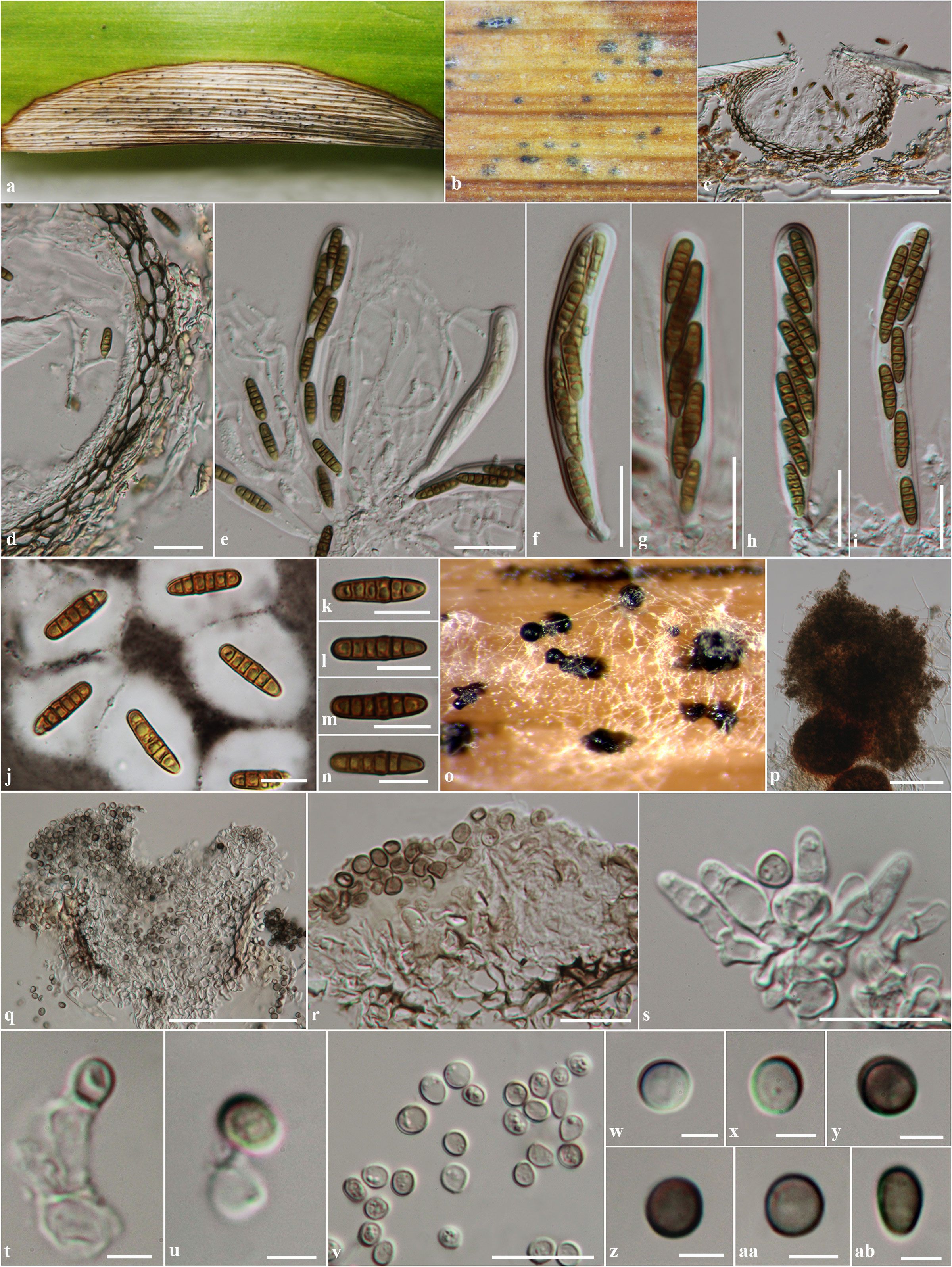
Fig. 1 Phaeosphaeriopsis dracaenicola (MFLU11-0193, holotype). on living leaves of Dracaena lourieri. a Leaf lesion on host. b Ascomata visible on host surface. c Section through ascoma. d Section through peridium. e Asci with pseudoparaphyses. f-i Asci. j Ascospores stained in Indian ink. k-n Ascospores. o Conidiomata forming on bamboo pieces on water agar. p Pycnidia. q Section through pycnidium. r Section through pycnidial wall. s-u Conidiogenous cells. v Conidia. w-ab Conidia. Scale bars: c = 100 µm, p, q = 50 µm, d, e, f, g, h, i = 20 µm, j, k, l, m, n, r, s, v = 10 µm, t, u, w, x, y, z, aa, ab = 2 µm.