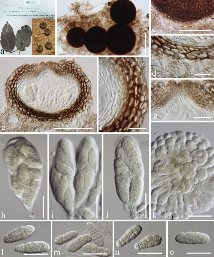
Phaeosaccardinula malloti (Rehm) Theiss., in Theissen & Sydow, Annls mycol. 15(6): 481 (1918) [1917], Figure 11
≡ Limacinula malloti Rehm, Philipp. J. Sci., C, Bot. 8(5): 395 (1913)
= Deslandesia paulensis var. malloti (Rehm) Bat. & Cif., Beih. Sydowia 3: 48 (1962)
MycoBank number:MB 156268; Index Fungorum number: IF 156268; Facesoffungi number: FoF 10341;
Epiphytic on the upper surface of living leaves of Mallotus philippensis, forming a sooty-like coating. Mycelium 4–6 μm wide (x̅ = 5.3 μm, n = 20), superficial, black, composed of dark brown to black, reticulate to branched, septate hyphae. Sexual morph: Ascomata 135–250 μm diam (x̅ = 220 μm, n = 10), superficial, scattered, globose to subglobose, cupulate when dry, dark brown to black, lacking setae, thick-walled, ostiolate. Ostiole in the center, periphysate or apapillate. Wall of ascoma 25–42 μm wide, multi-layered, externally comprising pigmented, dark brown, thick-walled cells of textura globulosa, with inner layer thinner, composed of lightly pigmented to hyaline, thin-walled cells of textura prismatica. Hamathecium lacking pseudoparaphyses. Asci 80–110× 18–32 μm (x̄ = 103 × 28 μm, n = 10), 8-spored, bitunicate, oblong-ellipsoid, broadly clavate, subglobose to oval when young, shortly pedicellate, early evanescent, lacking an ocular chamber when mature. Ascospores 18–24 × 6–9 μm (x̄ = 20 × 8 μm, n = 10), overlapping 2–4-seriate, hyaline, olivaceous-green at the septa of mature ascospores, oblong-ellipsoid, muriform, with 4–7 transverse septa and 4–6 longitudinal septa, constricted at the septa, rounded at both ends, with a mucilaginous sheath. Asexual morph: Undetermined.
Material examined: Philippines, Los Baños, on leaves of Mallotus philippensis (Lam.) Müll. Arg. (Euphorbiaceae), March 1913, C. F. Baker (PC0084486, holotype).

Figure 11 Phaeosaccardinula malloti (PC0084486, holotype). a Herbarium material, with superficial ascomata on the host. b, c Squash mounts of ascomata. d, f Vertical section through peridium. e Vertical sections of ascoma. g Vertical section through ostiole. h–k Asci with ascospores. l–o Ascospores. Scale bars: b = 200 µm, c, e =100 µm, d, f, g, h–j, l–o = 20 µm, k = 50 µm.
