Phaeonawawia diplocladielloidea Goh, J.H. Ou & C.H. Kuo, Mycol. Progr. 20(3): 228 (2021)
MycoBank number: MB 837328; Index Fungorum number: IF 837328; Facesoffungi number: FoF14616;
Etymology – From Greek, −oides (resembling), and the generic name Diplocladiella G. Arnaud, the epithet “diplocladielloidea” denotes that this fungus is similar to Diplocladiella in producing dematiaceous stauroconidia.
Colonies on natural substratum effuse, brown, somewhat glistening. Mycelium partly superficial, partly immersed in the substratum, consisting of smooth, brown branched, septate hyphae. Conidiophores absent or rudimentary in the form of a basal stalk (10–20 × 4–6 μm) under the single conidiogenous cell. Conidiogenous cells phialidic, discrete or integrated, sessile or incorporated terminally in the basal stalk, bulbose, doliiform or urceolate, (26.5)30–35.5 × 10–11.5 μm, with a distinct, rimmed opening (3.5–6 μm wide) at the apex, monoblastic. Sometimes regenerating percurrently. Conidia staurosporous, with 3–4 arms, broadly rounded or sometimes truncated at the tip of the arms, enteroblastic, exogenous, solitary, dry, smooth-walled, enclosed by a hyaline sheath (2–2.5 μm thick), tetrahedral to obpyramidal or occasionally ellipsoidal, mostly an equilateral triangle in surface view, with a length of (53)60–70(76.5) μm at each side and a height of 50– 61 μm, multicellular, arms 2–4-euseptate, septa thick and sometimes banded, not constricted at the septa, olivaceous to medium brown and versicoloured, with the central cell darker and the arms paler, each arm bears a hyaline, aseptate, filiform appendage (25–48.5 × 2–2.5 μm); ellipsoidal conidia 75– 85 × 25–30 μm, with two arms, each bearing a setula. Conidial secession schizolytic.
Teleomorph – Unknown
Conidiogenesis – Conidium ontogeny is enteroblastic, monoblastic, begins as a spherical, hyaline blown-out at the opening of the bulbose phialide. The blown-out then enlarged and becomes more or less ellipsoidal, positioning horizontally at the opening of the phialide, with its proximal side more tapering. The young conidium during the early development stages is hyaline and subsequently becomes more pigmented, while it continues to enlarge and finally becomes septate, setulate, and enclosed by a thick hyaline sheath. The conidium either develops into a horizontally oriented, ellipsoidal and bisetulate form, or into an obpyramidal to tetrahedral, 3–4- armed stauroform, with a hyaline setula at the end of each arm. Occasionally, the phialides may regenerate by forming a new phialide inside or outside the old ones (Fig. 3i). The process of conidiogenesis and phialide regeneration is depicted in Fig. 5.
Specimen examined – MALAYSIA. PERAK. Menglembu, Bukit Kledang, 4.58027–101.02528, 253 m a.s.l., on submerged wood, 9 January 2014, leg. Wai-Yip Lau and Teik- Khiang Goh, UTAR-G1, Herb. no. TNM: F0034163 (holotype), NCYU-UTAR-G1 (isotype).
GenBank Accession numbers – ITS: MT946684
Note – Phaeonawawia diplocladielloidea is unique in producing dematiaceous staurospores from discrete bulbose phialides. Its conidia resemble those of Diplocladiella species the most, especially those of D. alta R. Kirschner & Chee J. Chen; D. aquatica O.H.K. Lee, Goh & K.D. Hyde; D. cornitumida F.R. Barbosa, Gusmão & R.F. Castañeda; and D. tricladioides Nawawi, which are Y-shaped or triangular in face view, and bear filiform appendages at the end of the conidial arms. However, these species differ in having distinct conidiophores which are geniculate, cicatriced, with integrated polyblastic sympodial conidiogenous cells (Nawawi 1985b; Lee et al. 1998; Kirschner and Chen 2004; Barbosa et al. 2007). Two other fungi also have stauroconidia which are comparable with those of P. diplocladielloidea. In Jerainum triquetrum Nawawi & Kuthub., conidia are triangular in face view, muriform, bearing a single hyaline appendage at the base and several others at the distal ends. However, the conidiogenous cells are not phialides, instead, they are holoblastic, monotretic, and percurrently extending, although they are doliiform or ellipsoidal reminiscent of phialides (Nawawi and Kuthubutheen 1992). The stauroconidia of Triposporium elegans Corda, also resemble those of P. diplocladielloidea but lack f iliform appendages, and also differ in conidiogenesis, being holoblastic and non-phialidic (Corda 1837; Ellis et al. 1951). Several other hyphomycetes have discrete and/or integrated bulbose phialides similar to those of Phaenawawia, especially those of Craspedodidymum spp. (Yanna et al. 2000; Pinruan et al. 2004; Ma et al. 2011; Mel’nik et al. 2014), Obeliospora spp. (Nawawi and Kuthubutheen 1990; Wu and McKenzie 2003; Cantillo- Pérez et al. 2018), and Polybulbophiale palmicola (Goh and Hyde 1998b), but they all differ in conidial morphology.
Table 2 Comparison of hyphomycete genera similar to Phaeonawawia
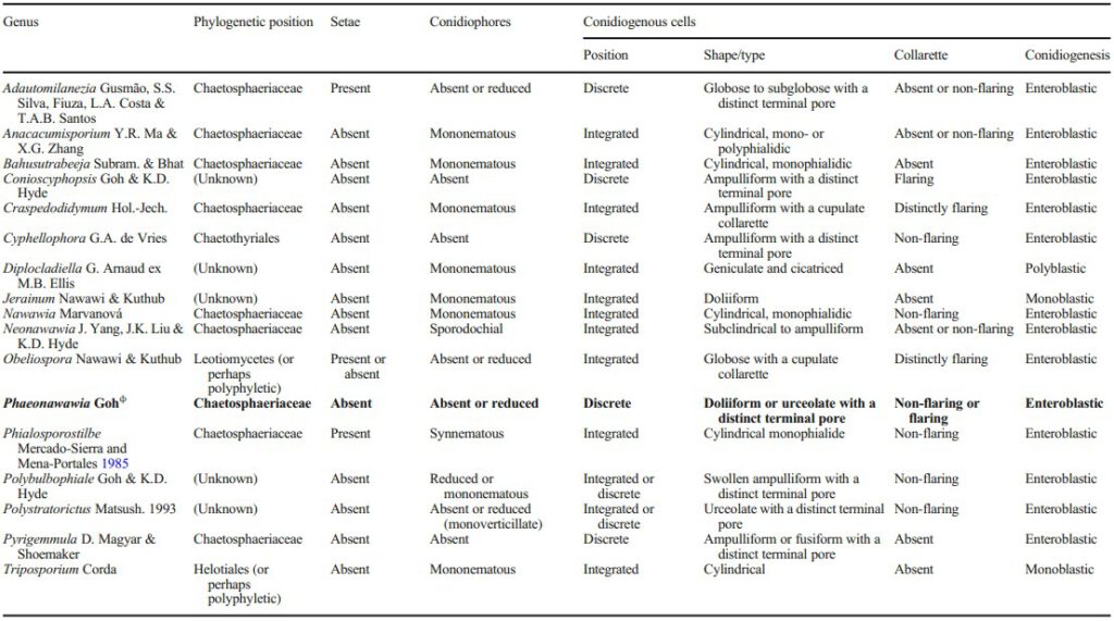
Table 2 (continued)
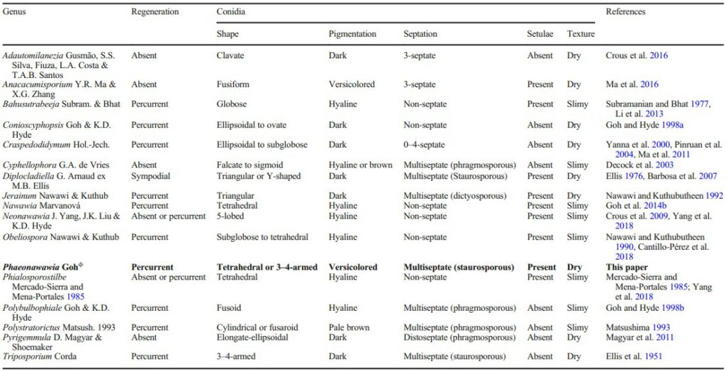
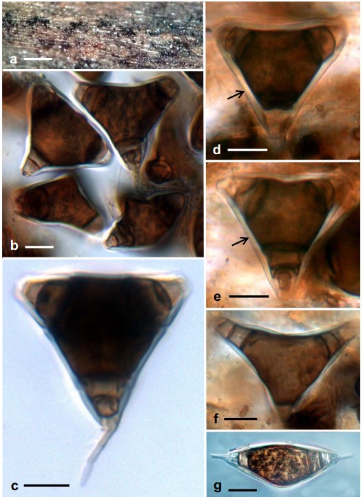
Fig. 1 Phaeonawawia diplocladielloidea (TNM: F0034163, holotype). a Colonies on the natural substratum (submerged wood). b–f Conidia, each with a thick hyaline sheath (arrowed in d and e). g An ellipsoidal conidium, bearing one hyaline appendage at each end. Scale bars: a = 500 μm, b–g = 20 μm
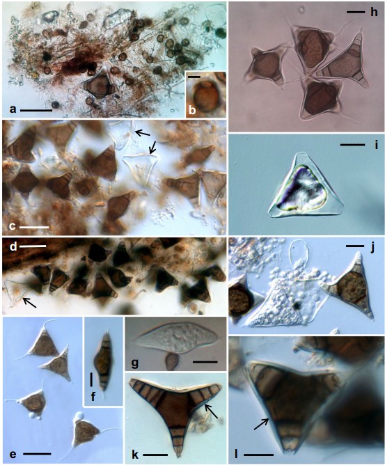
Fig. 2 Phaeonawawia diplocladielloidea (TNM: F0034163, holotype). a Squashed mount from the natural substratum showing a stauroconidium and many bulbose conidiogenous cells. b Close-up of a bulbose conidiogenous cell with a terminal opening. c, d Squashed mount of conidia. Arrows point to empty hyaline conidial sheaths. e Four conidia bearing hyaline filiform appendages, one at each arm. f An ellipsoidal conidium, bearing one hyaline appendage at each end. g A developing conidium at the opening of a bulbose conidiogenous cell. h Four tetrahedral or obpyramidal conidia bearing hyaline filiform appendages. i An empty conidial sheath. j Conidia beside empty conidial sheaths. k, l Conidia. Arrows point to a thick hyaline sheath enclosing the conidium. Scale bars: a, c–e = 50 μm, f–l = 20 μm, b = 5 μm
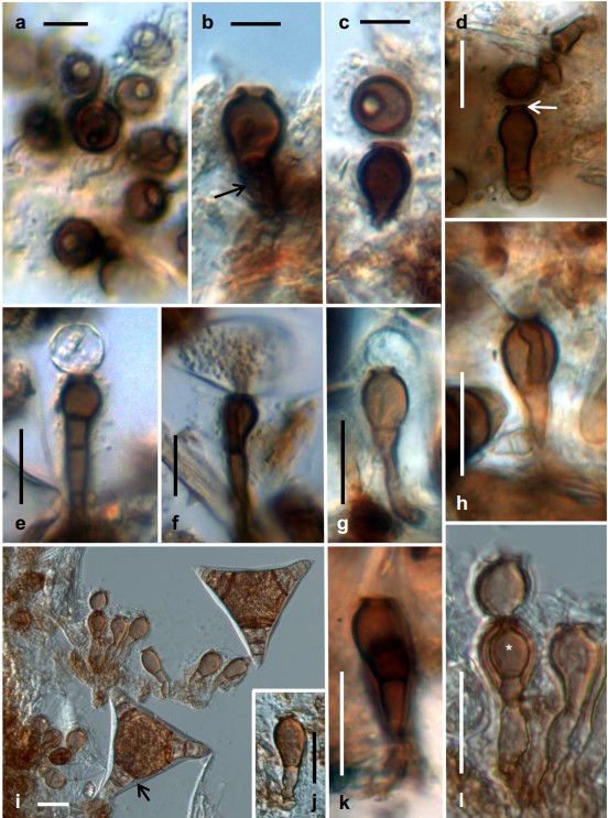
Fig. 3 Phaeonawawia diplocladielloidea (TNM: F0034163, holotype). a Several bulbose conidiogenous cells, each with a terminal opening. b Vertical view of a conidiogenous cell with a flared collarette at the terminal opening. Arrow points to the short basal stalk. c Two bulbose conidiogenous cells, one view from the top and the other from the side. d Vertical view of a conidiogenous cell with a short neck and flared collarette (arrowed) at the terminal opening. e–h Developing conidia at the opening of conidiogenous cells. i Two conidia and several stalked conidiogenous cells. Arrow points to the hyaline conidial sheath. j A stalked conidiogenous cell. k, l Stalked conidiogenous cells showing percurrent regenerations (highlighted by an asterisk in l). Scale bars: a–c = 10 μm, d–l = 20 μm
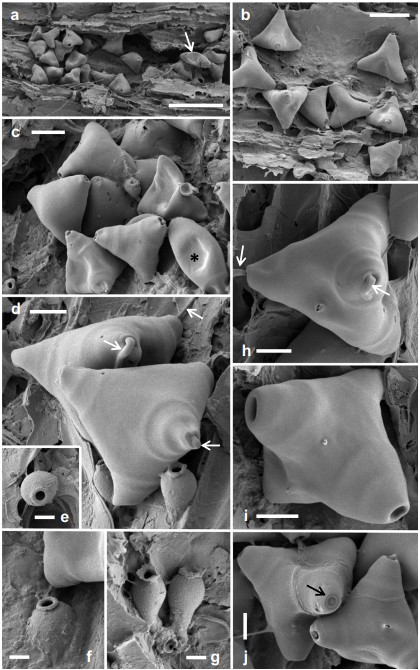
Fig. 4 Phaeonawawia diplocladielloidea (TNM: F0034163, holotype). Scanning electron micrographs. a Colony on the natural substratum. Arrows point to an ellipsoidal conidium. b, c Clumps of conidia. The asterisk denotes an ellipsoidal conidium. d Two tetrahedral (obpyramidal) conidia and two conidiogenous cells. Filiform appendages are visible (arrowed). e–g Conidiogenous cells. h–j Conidia. Arrows in h point to filiform appendages. Large pores in i are ends of conidial arms lacking filiform appendages. Arrow in j points to the basal hilum of an obpyramidal conidium. Scale bars: a = 100 μm; b = 50 μm; c = 20 μm; d, h–j = 10 μm, e-g = 5 μm
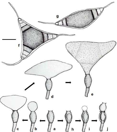
Fig. 5 Phaeonawawia diplocladielloidea. Diagrammatic representation of conidiogenesis. a A discrete phialide situated on a basal stalk. b–e Sequential steps of conidiogenesis. f A mature stauroconidium bearing a setula at the end of each arm. g A mature ellipsoidal conidium bearing a setula at each end. h An old phialide showing percurrent regeneration. i Conidium ontogeny from regenerated phialide. j Percurrent regeneration of phialides. Scale bar = 20 μm
