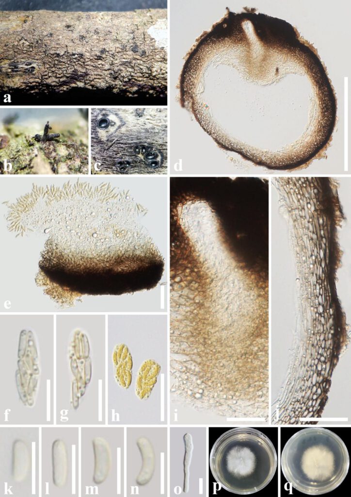
Peroneutypa aquilariae T.Y. Du & Tibpromma, sp. nov. (Fig. 2)
MycoBank number: MB 845438; Facesoffungi number: FoF 12744
Etymology – named after the host genus, Aquilaria.
Saprobic on dead twigs of Aquilaria sinensis (Thymelaeaceae). Sexual morph: Ascostromata 0.5–1.5 mm wide, well-developed interior, solitary to gregarious, mostly solitary, immersed, long ostiolar canal obviously raised through host tissue, black, irregular in shape, arranged irregularly, 1–6 locules. Ascomata (excluding necks) 300–570 μm diam., perithecial, immersed in ascostromata, subglobose to globose, dark brown to black. Ostiolar canal 20–40 μm wide, without paraphyse, filled with hyaline cells, with 200–300 μm long, cylindrical, straight, dark-brown to black necks. Peridium 35–70 μm wide, composed of two layers, outer layer comprising several layers, thick-walled, dark brown to pale brown cells of textura angularis, inner layer comprising 3–5 layers, thin-walled, hyaline cells of textura prismatica. Paraphyses absent. Asci 15–20 × 5–7 µm (x̄ = 18.3 × 5.8 μm, n = 20), unitunicate, 8-spored, clavate to cylindrical, thin-walled, short or without pedicellate, apically rounded to truncate with indistinct J-apical ring. Ascospores (5–)5.5–7(–7.5) × (1.6–)1.8–2.2 µm (x̄ = 6.3 × 2 μm, n = 30), overlapping 1–3-seriate, hyaline to pale yellow, oblong to allantoid, slightly curved, aseptate, smooth-walled, with small guttules, ascospores turn to yellow after being stained by Melzer’s reagent. Asexual morph: Undetermined.
Culture characteristics – Ascospores germinated on PDA within 24 h at a constant temperature incubator (28 ℃). Colonies on PDA reaching 6 cm diam., after one week at 28 ˚C, mycelium white, flossy, circular with the entire edge, with filiform margin. After one month, mycelium becomes white to light yellow above, and light brown to brown in reverse.
Material examined – CHINA, Yunnan Province, Xishuangbanna, on dead twigs of Aquilaria sinensis (Thymelaeaceae), 15 September 2021, Tianye Du, YNA3 (holotype, HKAS 124185; ex-type cultures, KUNCC 22-10817; ex-isotype, KUNCC 22-10818).
Notes – Phylogenetic analyses of combined ITS and tub2 showed Peroneutypa aquilariae is well separated from other strains in Peroneutypa (Fig. 1). Peroneutypa mackenziei (MFLUCC 16-0072) is phylogenetically closely related to P. aquilariae, however, they differ in morphological characteristics i.e. stromata of Peroneutypa aquilariae has well-developed interior, ostiolar canal without periphyse, peridium inner layer comprises of 3–5 hyaline cell layer of textura prismatica, and paraphyses absent; while P. mackenziei has poorly developed interior stromata, ostiolar canal periphysate, peridium inner layer comprises of 8–10 hyaline cell layer of textura angularis, and hamathecium composed paraphyses. In addition, the asci and ascospores of Peroneutypa aquilariae are slightly larger than those of P. mackenziei (asci 18.3 × 5.8 µm vs 17.7 × 4.2 µm; ascospores 6.3 × 2 µm vs 5.6 × 1.6 µm) (Shang et al. 2017). Table 3 shows more information about countries, hosts, and a comparison of morphological characteristics of Peroneutypa. Based on both morphological characteristics and multigene phylogenetic analyses results, we introduce Peroneutypa aquilariae as a distinct new species.

Fig. 2 – Peroneutypa aquilariae (HKAS 124185, holotype). a-c. Appearance of ascomata on the substrate. d. Section through ascoma. e-g. Asci. h. Asci stained by Melzer’s reagent. i. Ostiole. j. Peridium. k-n. Ascospores. o. A germinating ascospore. p, q. Colony on PDA medium (after 7 days in culture). – Scale bars: d = 300 μm, e, j = 50 μm, f, g = 10 μm, h = 20 μm, i=30 μm, k-o=5μm.
