Periconia pseudobyssoides Markovsk. & A. Kačergius, Mycol. Progr. 13(2): 293 (2013) [2014]
MycoBank number: MB 804763; Index Fungorum number: IF 804763; Facesoffungi number: FoF 03857; Fig. **
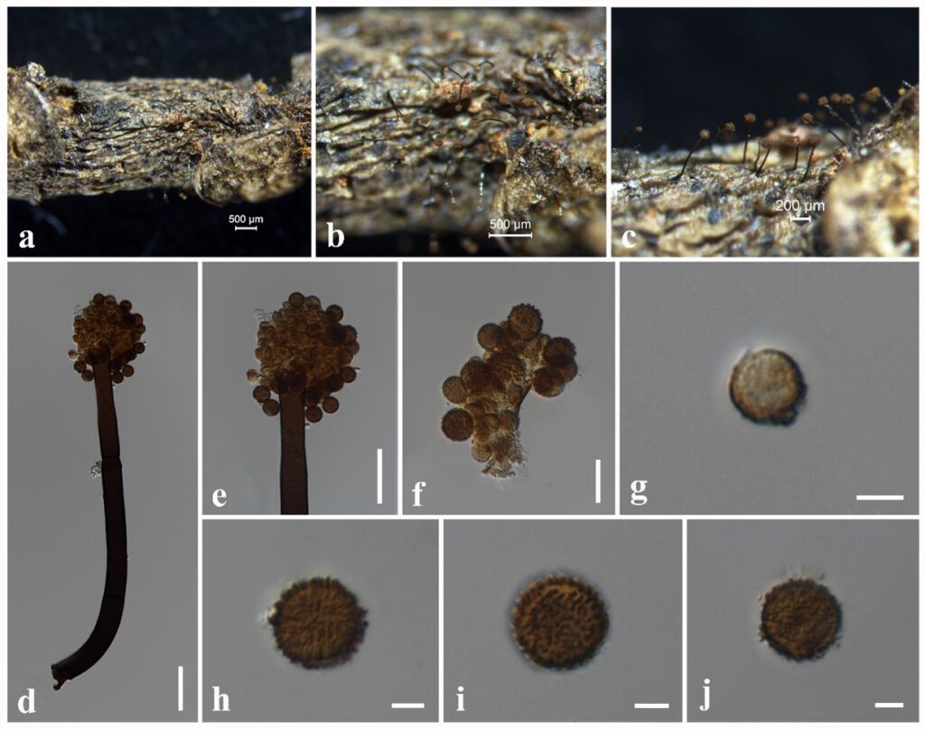
Saprobic on dead twigs attach to Magnolia sp. Sexual morph: Undetermined. Asexual morph: Hyphomycetous. Colonies on substrate numerous, effuse, dark brown to black, floccose. Conidiophores 350–370 × 22–25 μm ( = 360 × 23 µm, n = 10), macronematous, mononematous, unbranched, erect, straight or slightly flexuous, single, light brown to dark brown, septate, thick-walled. Conidia 10–15 × 10–15 μm ( = 14 × 14 µm, n = 30), solitary, globose, light brown to dark brown, finely verruculose, aseptate.
Culture characteristics – Colonies on PDA reaching 30 mm diameter after 1 weeks at 25°C, colonies from above: circular, margin entire, dense, slightly raised, fairy fluffy appearance, cream; reverse: pale brown.
Material examined – CHINA, Yunnan Province, Xishuangbanna, dead twigs attached to Magnolia sp. (Magnoliaceae), 27 March 2017, N. I. de Silva, NI273 (MFLU 18-2651), living culture, MFLUCC 19-0041.
Known hosts and distribution – On dead stalks of Heracleum sosnowskyi in Lithuania (Markovskaja & Kačergius 2014), on dead twigs of Magnolia sp. in China (this study).
GenBank numbers – ITS: OL966949, LSU: OL830815, tef1: *******.
Notes – Periconia pseudobyssoides was introduced by Markovskaja & Kačergius (2014) from dead stalks of Heracleum sosnowskyi in Lithuania. The morphology of our collection (MFLU ******) shares similarities with Periconia pseudobyssoides in having macronematous, mononematous, unbranched, light brown to dark brown, septate conidiophores and globose, light brown to dark brown, asptate conidia (10–15 × 10–15 μm vs 15–17 μm diam.) (Markovskaja & Kačergius 2014). Multi-gene phylogeny also indicates that our collection clusters with other Periconia pseudobyssoides isolates in 82% ML, 1.00 BYPP supported clade (Fig. **). Therefore, we introduce our collection as a new host record of Periconia pseudobyssoides from dead twigs of Magnolia sp. in Thailand.

Figure * – Periconia pseudobyssoides (MFLU 18-2651) a The specimen. b, c Appearance of colonies on substrate. d Conidiophore with conidia. e Part of conidiophore with conidia. f Conidiogenesis cells and conidia. g–j Conidia. Scale bars: a, b = 500 μm, c = 200 μm, d = 50 μm, e, f = 20 μm, g–j = 5 μm.
