Pedrocrousiella pongamiae (Syd. & P. Syd.) Rajeshkumar, U. Braun & J.Z. Groenew., comb. nov.
MycoBank number: MB 838147; Index Fungorum number: IF 838147; Facesoffungi number: FoF 11193; Figs 2–5.
Basionym: Fusicladium pongamiae Syd. & P. Syd., Ann. Mycol. 11(4): 328. 1913.
Synonyms: Passalora pongamiae (Syd. & P. Syd.) Subram., Hyphomycetes: 237. 1971.
Asperisporium pongamiae (Syd. & P. Syd.) Deighton, In Ellis, More Dematiaceous Hyphomycetes: 241. 1976.
Asperisporium pongamiae-pinnatae Khawar, A. Kumar, Bhat & C. Nakash., Vegetos 25(1): 336. 2012.
Typus: India, Tamil Nadu, Coimbatore, Iruttu Palam, on leaves of Pongamia pinnata (= P. glabra), Dec. 1909, C.E.C. Fischer (lectotype designated here, S-F45748, MycoBank MBT395139). India, Maharashtra, Pune, Kothrud, on leaves of Pongamia pinnata, 23 Jun. 2018, K.C. Rajeshkumar (epitype designated here AMH 10302, MycoBank MBT395140); culture ex- epitype NFCCI 4881. Ex-epitype sequences: MW327548 (ITS), MW327593 (LSU), MW363496 (rpb2).
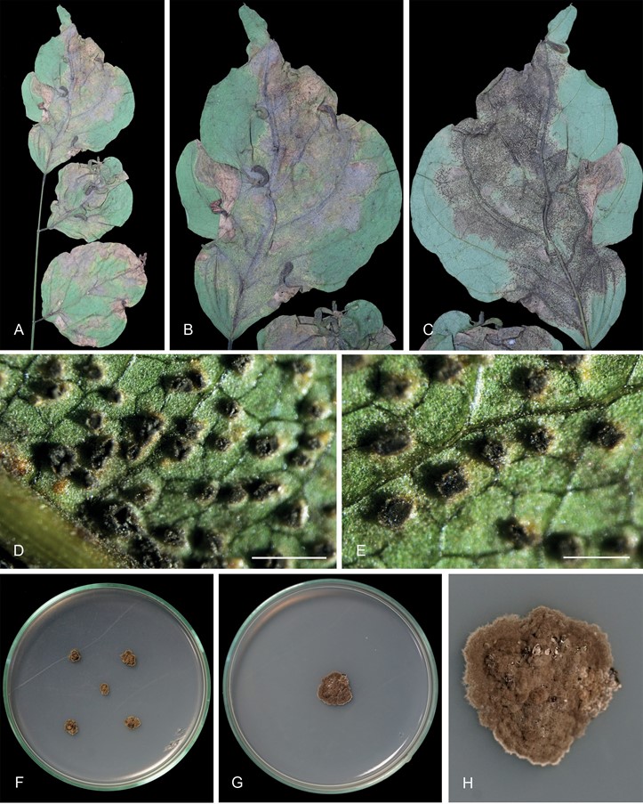
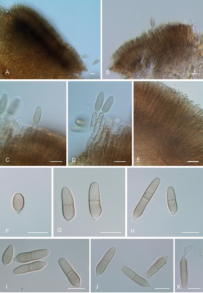
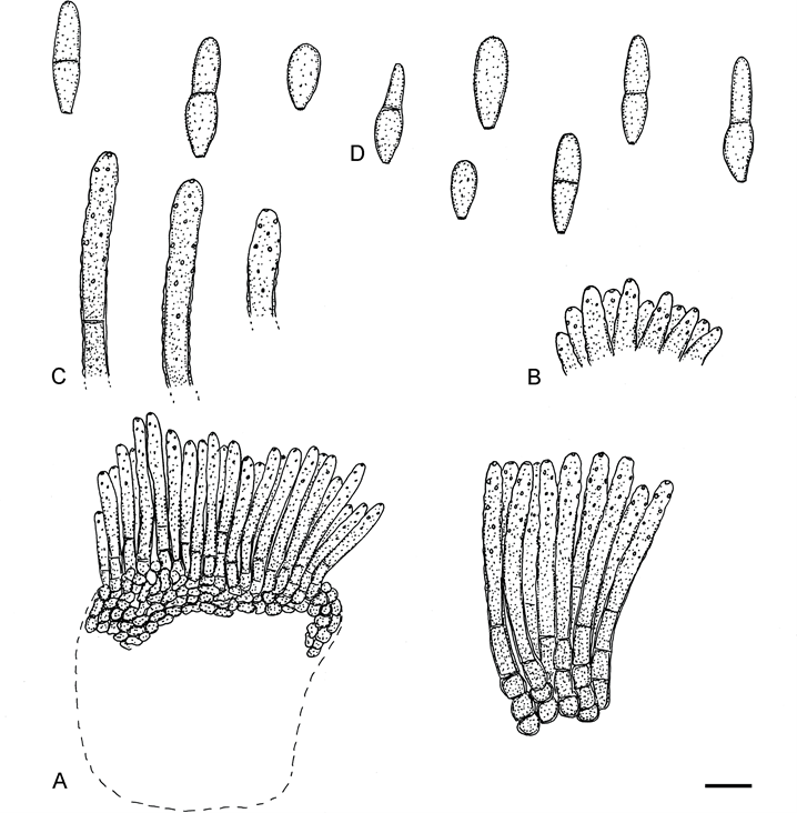
In vivo: Phytopathogenic, causing leaf spots, amphigenous, colour, shape and size variable, subcircular to irregular, sometimes diffuse, 2–20 mm diam or confluent and larger, sometimes large leaf segments or almost entire leaves discoloured, yellowish green, ochraceous to brownish, reddish brown, later becoming dark brown to blackish brown by the development of abundant sporodochia. Mycelium internal. Stromata immersed, well-developed, large, to 120 µm diam, medium to dark brown, composed of swollen hyphal cells, 2–7 µm diam, rounded in outline to irregularly shaped. Conidiomata sporodochial, mostly hypophyllous, scattered to usually dense, olive brown, dark brown to blackish, erumpent, 50–250 μm diam. Conidiophores densely fasciculate, very numerous, arising from stromata, macronematous, mononematous, septate below or conidiophores reduced to conidiogenous cells, unbranched, straight to slightly sinuous, but not geniculate, smooth to verruculose-rugose, pale brown, wall thin or somewhat thick- walled, 35–65 × 2.5–5 μm. Conidiogenous cells integrated, terminal, up to about 40 µm long, cylindrical, proliferation sympodial, but not causing any trace of geniculation, polyblastic, usually with numerous, often densely arranged thickened and darkened conidiogenous loci, 1–2 µm wide. Conidia formed singly, broad ellipsoid, ovoid, short subcylindrical or obclavate, apices obtuse, bases truncate, 15–30 × 4.5–7 μm, young conidia 10–12.5 × 5–5.5 μm, 0–1(–2)-septate, wall pale olivaceous to olivaceous brown, thin, verruculose, basal hilum slightly thickened and darkened or almost undifferentiated, 0.5–1.5 μm wide, schizolytic.
Colonies on MEA at 25 ± 2 °C after 15 d slow growing, 20–28 mm (40–48 mm after 45 d) diam, dark brown to black, velutinous with umbonate centre, reverse black. Colonies on CMA at 25 ± 2 °C after 15 d 20–25 mm, velutinous, blackish, reverse black.
Additional materials examined: India, Tamil Nadu, former Madras Presidency, Malabar District, Chalisseri, on leaves of Pongamia pinnata, 10 Jul. 1912, W. McRay (S-F45747, syntype); Maharashtra, Pune, Kothrud, on leaves of Pongamia pinnata, 9 Sep. 2020, K.C. Rajeshkumar RKC-2020; cultures RKC-2020.1 and RKC-2020.2. Sri Lanka, Peradeniya, on leaves of Pongamia pinnata, Dec. 1913, T. Petch [Petrak, Mycoth. Gen. 736] (M); ibid. [Syd., Fungi Exot. Exs. 441] (M).
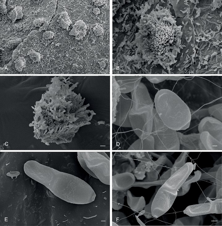
Notes: Scanning Electron Microscopy studies confirmed the erumpent sporodochial nature of the conidiomata, the densely fasciculate conidiophores and ovoid to obclavate conidia having verruculose conidial ornamentation as observed by light microscopy. Asperisporium pongamiae-pinnatae (Khawar et al. 2012) is undoubted a heterotypic synonym of A. pongamiae. Type material of A. pongamiae-pinnatae was not available for re- examination, but this species shares Pongamia pinnata as type host with A. pongamiae and, based on its original description, it is morphologically indistinguishable from A. pongamiae.

Fig. 2. Pedrocrousiella pongamiae (AMH 10302 – epitype). A–C. Symptoms on the upper and lower surface of the host leaves, Pongamia pinnata. D, E. Sporodochial development on abaxial surface. F. Colonies on MEA after 15 d. G, H. Colonies on MEA after 45 d. Scale bars = 500 µm.

Fig. 3. Pedrocrousiella pongamiae (AMH 10302 – epitype). A, B. Scanning Electron Micrograph of sporodochia on abaxial surface of leaves. C. Section through sporodochia showing conidiophores. D. Young ovoid detached conidium with truncate base. E, F. Mature obclavate conidia. Scale bars: A, B = 20 µm, C = 10 µm, D–F = 1 µm.

Fig. 4. Pedrocrousiella pongamiae (AMH 10302 – epitype). A, B. Sporodochia. C, D. Conidiophores with cicatrised conidiogenous cells and attached conidia. E. Densely fasciculate conidiophores. F. Young broad, ellipsoid, aseptate conidium. G, H. Mature, obclavate, 1–2-septate conidia. I–K. Conidial variation. Scale bars = 10 µm.

Fig. 5. Pedrocrousiella pongamiae (Syd., Fungi Exot. Exs. 441 – M). A. Section through a sporodochium. B. Fasciculate conidiophores. C. Apical part of conidiophores. D. Conidia. Scale bar = 10 µm. U. Braun del.
