Parascedosporium putredinis (Corda) Lackner & de Hoog
Saprobic or pathogenic on plant material, or isolated from soil and dung. Sexual morph: Undetermined. Asexual morph from the natural substrate: Conidiomata graphium-like synnema. Synnemata 707–851 μm tall, erect, with a cylindrical stipe. Stipe 8–14 μm wide, olivaceous grey, smooth-walled, terminating in a slimy head of conidia, appearing as a slimy droplet, 31–64 μm. Conidiogenous cells 12.8–21 × 1.1–1.9 μm (x̅ = 15.3 × 1.4 μm; n = 10), percurrent, cylindrical, smooth-walled. Conidia 4.2–6.1 × 2.4–3.4 μm (x̅ = 5 × 2.8 μm; n = 25), oblong, hyaline, smooth-walled. Synnematal stage from the culture: Conidiomata graphium-like synnema. Synnemata 300–555 μm tall, erect, with a cylindrical stipe. Stipe 28–41 μm wide, dark grey, smooth-walled, terminating in a slimy head of conidia, appearing as a slimy droplet, 83–255 μm. Conidiogenous cells 17–22 × 0.8–1.8 μm (x̅ = 20.3 × 1.3 μm; n = 10), percurrent, cylindrical, hyaline, smooth-walled. Conidia 6.8–9.2×3–4.5 μm (x̅ = 8 × 3.8 μm; n = 25), ovate to subcylindrical, base obtuse to subtruncate, aseptate, straight, multi-guttulate, hyaline, smooth-walled. Solitary conidiophores from culture: Conidiophores 1.5–2.1 μm wide, solitary, emerging from aerial mycelium, undifferentiated, simple, often reduced to conidiogenous cells, or irregularly branched, with branches often bearing two to five conidiogenous cells, thin-walled. Conidiogenous cells 8.6–24.4 × 1.7–2.5 μm (x̅ = 14.1 × 2.2 μm; n = 20), cylindrical to flask-shaped, hyaline, thin-walled, denticulate, usually terminating in a cluster of two to three cylindrical denticles. Conidia 4.7–7.3 × 2.7–4.2 μm (x̅ = 6.2 × 3.5 μm; n = 25), obovate or subglobose, hyaline, smooth, more or less thick-walled.
Culture characters: Reaching 20–27 mm within 7 days on MEA, at 25 °C, circular, with sparse aerial mycelium, and smooth, margins lobed, white from above, reverse gray in the center, becoming white towards the margin. Aerial mycelium produced simple conidiophores within 7 days at the margins. Synnemata produced on MEA within 30 days. Colonies becoming dark gray, and slightly granular at the centre due to the abundance of synnemata.
Material examined: THAILAND, Chiang Rai Province, Mae Fah Luang University campus, on a dried seed pod of Delonix regia (Boj. ex Hook.) Raf. (Fabaceae), 14 December 2015, R.H. Perera, RHP 130 (MFLU 17-0736), dry culture on MEA, MFLU 18-0516; living culture, MFLUCC 15-1009. GenBank: MH048678 (ITS), MH048679 (LSU), MH048680 (SSU), MH048681 (tef).
Notes: Our new isolate (MFLUCC 15-1009) clustered with other Parascedosporium putredinis strains in the phylogenetic tree. Differences of two bases were noted in the ITS region between our new isolate and the ex-type (CBS 102083). The new collection produced synnemata and lateral solitary conidiophores (in culture) similar to the holotype of P. putredinis (Figs 2–4). Our new P. putredinis isolate is slightly different from the holotype in possessing larger conidia (6.8–9.2×3–4.5 vs. 5.5–7.5×2.5–3.5 μm) (Gilgado et al. 2007). By considering the morphological and molecular data we designate our new collection as P. putredinis.
Fig. 1 Parascedosporium putredinis (MFLU 17-0736). a Herbarium material. b Conidiomata on the host substrate. c Conidiophore with conidia. d, e Close up of the apex of the stipe. f, g Conidiogenous cells with conidia. h Conidia. i Germinating conidia. j, k Colonies on MEA. Scale bars: b, c = 500 μm, d = 100 μm, e = 50 μm, f = 20 μm, g–i = 10 μm.
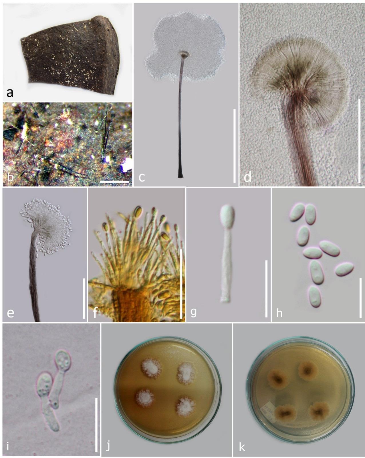
Fig. 2 Parascedosporium putredinis graphium stage on MEA (MFLU 18-0516). a, b Synnemata on MEA. c Synnemata. d Conidiophores with conidia. e Conidia. Scale bars: a = 1 mm, b = 500 μm, c = 200 μm, d, e = 20 μm.
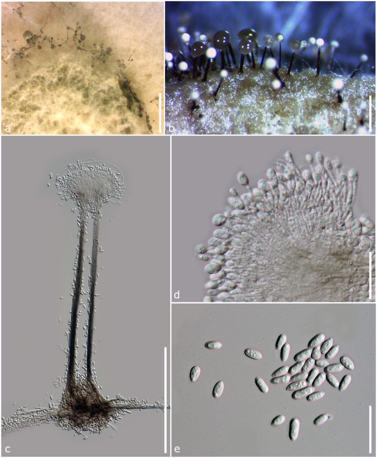
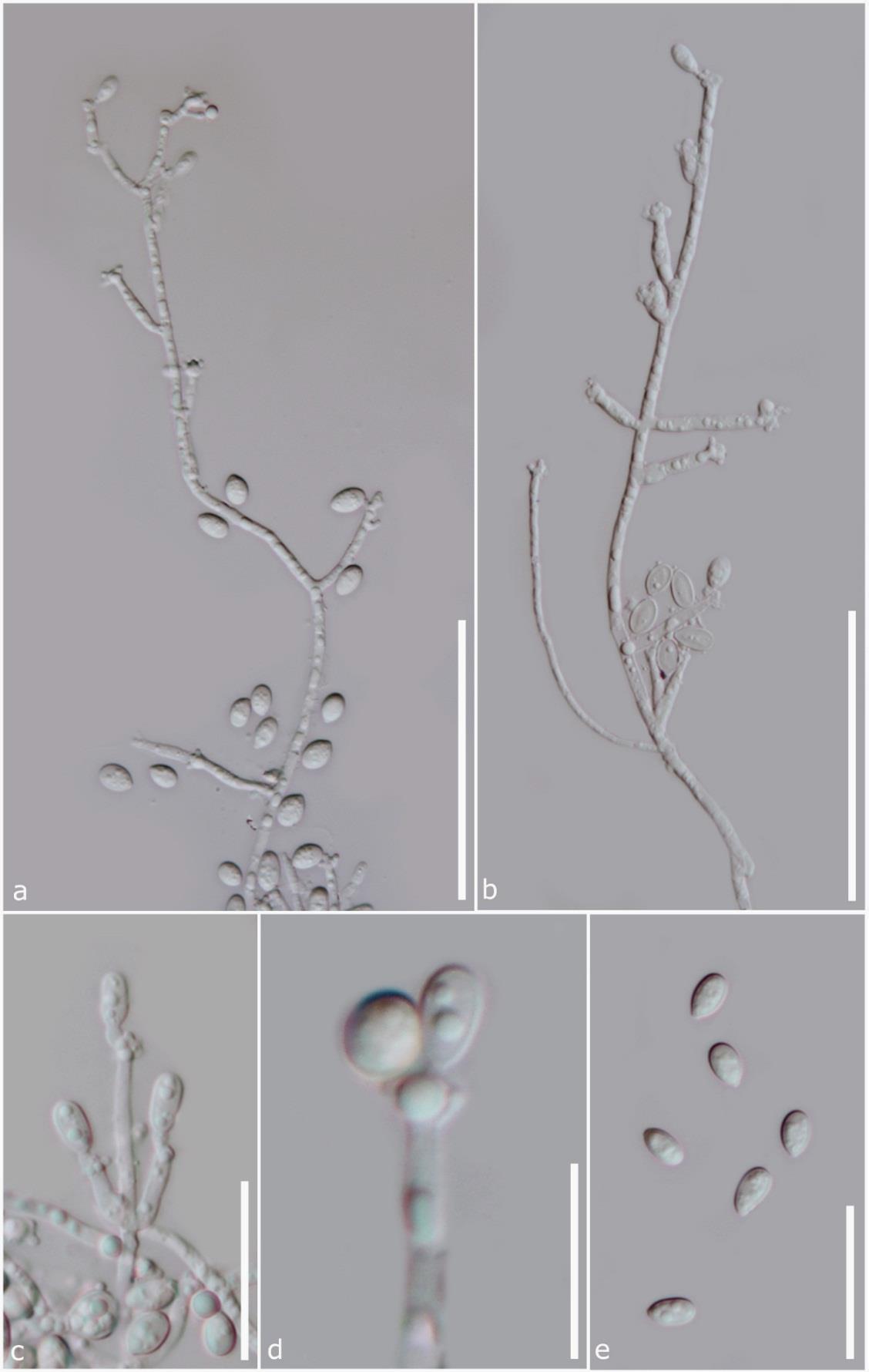
Fig. 3 Parascedosporium putredinis solitary conidiophores on MEA (MFLU 18-0516). a, b Aerial hyphae with simple or branched conidiophores producing sympodial conidia. c, d Lateral, cylindrical conidiogenous cells with conidia. e Conidia. Scale bars: a, b = 50 μm, c = 20 μm, d = 10 μm, e = 20 μm.