Parametarhizium S. Gao, W. Meng, Li Xiang Zhang, Q. Yue, L. J. Xu, gen. nov.
MycoBank number: MB 837521; Index Fungorum number: IF 837521; Facesoffungi number: FoF;
Etymology: based on its close morphological relationship to Metarhizium.
Type species: Parametarhizium hingganense S. Gao, W. Meng, Li Xiang Zhang, Q. Yue, L. J. Xu, sp. nov.
Diagnosis: Colonies white to yellow, velvety, exudate lemon yellow, sometimes radially sulcate, reverse pale yellow to brown. Conidiophores hyaline, arising from branches of aerial hyphae, with candelabrum-like arrangement of phialides. Phialides, 2– 4 in whorls, cylindrical to obpyriform. Conidia subglobose to ellipsoidal, hyaline (1.1–) 1.2–2.2 (–2.8) (1.0–) 1.6–1.7 (–2.6) µ m.
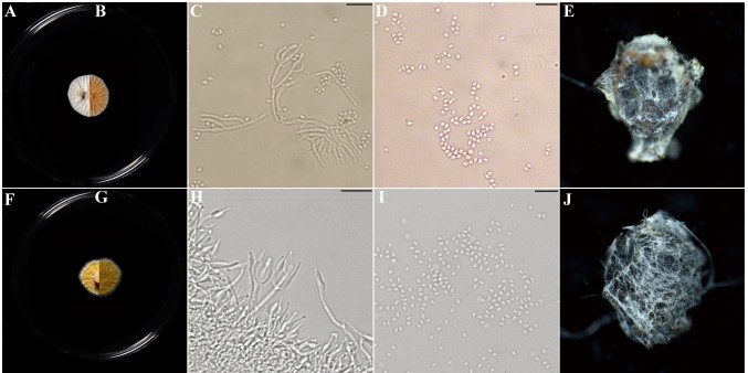
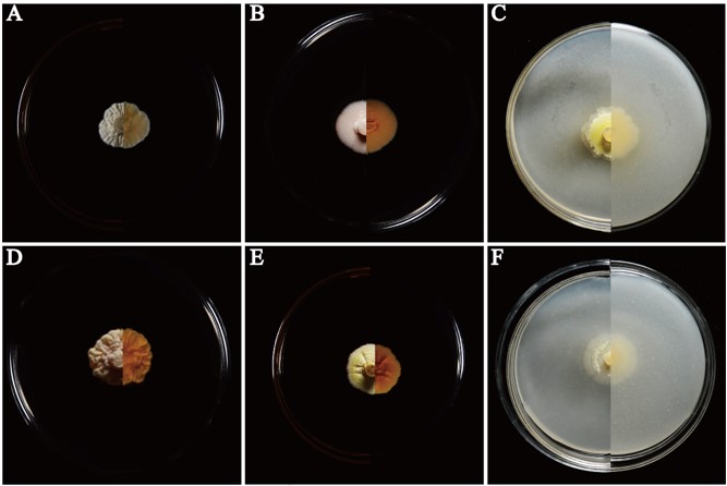
Description: Colonies on PDA, SDAY, MEA, OA, white to yellow, never green, radially sulcate, 25◦C reaching 13– 20 mm in 2 weeks, velvety; exudate lemon yellow; reverse pale yellow to brown. Hyphae hyaline, septate, smooth-walled, 0.8– 2.5 µm wide. Conidiophores hyaline, smooth-walled (20–) 34–62 (–100) (0.9–) 1.5–1.9 (–2.2) µm, arising from branches of aerial hyphae, with whorls of 2–4 phialides. Phialides candelabrum- like arrangement (2.7–) 5.6–17.5 (–26) (1.2–) 1.4–2.3 (–2.5) µm, cylindrical to obpyriform. Conidia subglobose to ellipsoidal, hyaline to yellow (1.1–) 1.2–2.2 (–2.8) (1.0–) 1.6–1.7 (–2.6) µ m (Figure 4 and Supplementary Figure S2).
Notes: – Compared with those of Metarhizium, the colonies of Parametarhizium are white to yellow (vs. green), and its subglobose-to-ellipsoidal conidia are smaller (<3.3 µm) than most of Metarhizium spp. Parametarhizium exhibits a candelabrum-like arrangement of phialides, while Metapochonia and Pochonia have Verticillium-like conidiophores, and other related genera show Nomuraea-like, Paecilomyces-like or Lecanicillium-like conidiophores.

FIGURE 4 | Morphological characters of Parametarhizium changbaiense [from ex-holotype China General Microbiological Culture Collection Center (CGMCC) 19143] and Parametarhizium hingganense (from ex-holotype CGMCC 19144). (A,B) The front and reverse of a P. changbaiense colony on PDA after 14 days at 25◦C. (C,D) The phialides and conidia of P. changbaiense. (E) Rhopalosiphum maidis infected by P. changbaiense. (F,G) The front and reverse of a P. hingganense colony on PDA after 14 days at 25◦C. (H,I) The phialides and conidia of P. hingganense. (J) R. maidis infected by P. hingganense.

Figure S2. Colonies of Parametarhizium changbaiense and Parametarhizium hingganense on different media. (A-C) P. changbaiense colonies (front left side, reverse right side) on Sabouraud dextrose agar with yeast extract (SDAY), Malt extract powder (MEA), and Oatmeal agar (OA). (D-F) P. hingganense colonies (front left side, reverse right side) on SDAY, MEA, and OA.
Species
