Parafuscosporella garethii Boonyuen, Chuaseehar. & Somrith., sp. nov., Index Fungorum Number: IF552573
Etymology: Named after Professor E.B. Gareth Jones in recognition and celebration of his 80th birthday in January 2017 and his contributions to tremendous changes in Asian fungal classification, identification and nomenclature, particularly on freshwater fungi in Thailand.
Saprobic on submerged wood. Colonies on natural substrata, granular, black. Mycelium mostly superficial and partly immersed in the substrata, composed of branched, septate, smooth-walled, hyaline, 1.25–2.5 μm thick hypha. Sexual morph: Undetermined. Asexual morph coelomycetous, sporidochial. Conidiomata sporodochial, scattered, spherical to cushion-shaped, with jelly-like cover, gelatinous, 0.5–0.8 × 0.5–0.7 mm diameter. Conidiophores micronematous, mononematous, compact, erect or flexuous, branched, septate, smooth-walled, hyaline, cylindrical or mostly moniliform with globose to subglobose or ellipsoidal to clavate cells, 10–15 × 7.5–8 μm, connected centrically, up to 62.5 μm long. Conidiogenous cells holoblastic, monoblastic, integrate or discrete, smooth-walled, hyaline, cylindrical, 1.25–2.5 μm wide or globose to subglobose, 8–12.5 μm wide, or ellipsoidal, 10–15 × 7.5–8 μm. Conidial secession rhexolytic. Conidia solitary, acrogenous, smooth-walled, obpyramidal, coronate at the apex, 2 (-3) celled with transverse septum near the base, distal cell black, basal cell (s) light brown, (37.5–) 40–47.5 × (25–) 27.5–42.5 μm, 20–32.5 μm thick (x̅ = 43.1 × 35.1 × 24.3 μm, n = 50), with a distinct basal frill 2–5 μm long. Conical projections arising from the tip of conidia, 4–9-conical, 5–7.5 × 5 μm.
Culture characteristics: Conidia germinating on PDA within 48 hours. Colonies on PDA slow-growing, attaining 12.6–15 mm diameter in 40 days at 20-25°C, floccose, rounded, greenish-grey at first, becoming grey to dark grey when aged (27F2/27F1); reverse sallow, yellowish-grey to dark grey with age (colours: 2B2/2F1; Kornerup & Wanscher JH 1963), mycelium partly immersed and partly superficial, sporulating with prolonged incubation time. Conidiophores reduced to conidiogenous cells. Conidiogenous cells holoblastic, monoblastic, integrate, cylindrical, smooth-walled, hyaline, 2–2.5 μm wide. Conidial secession rhexolytic. Conidia acrogenous, solitary, smooth-walled, obovoid to pyriform, 2(-3)-celled with transverse septum near the base, 22.5–30 × 15–25 μm (x̅ = 25.3 × 21.3 μm, n = 30), upper cell(s) brown to dark brown, basal cell light brown; Chlamydospores absent.
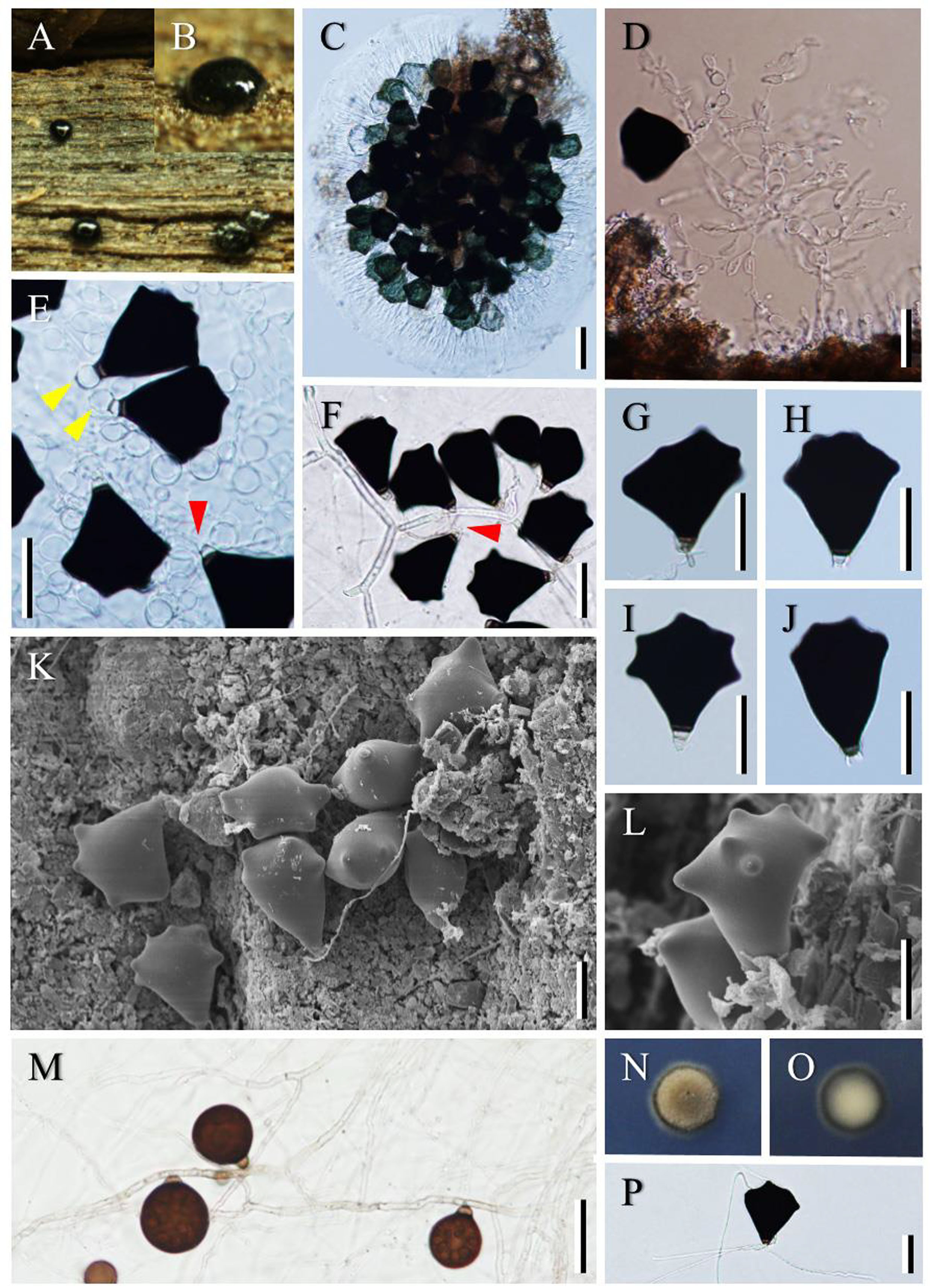
Fig. Parafuscosporella garethii (BBH 40839). A–B Sporodochia on submerged wood. C Squash mount of a sporodochium. D Conidiophores with conidiogenous cells and conidia. E–F Conidia with conidiogenous cells using yellow and red arrows showing conidia with globose to subglobose conidiogenous cells, and cylindrical conidiogenous cells, respectively. G–J Detached mature conidia. K–L Conidiophores, conidiogenous cells and conidia (SEM). M Sporulating conidia and its hyphae on PDA. N–O Obverse and reverse views of colony after incubated at 25°C on PDA for 30 days. P Germinating conidium on PDA. – Bars C = 50 μm; D–M, P = 25 μm.