Paradendryphiella salina (G.K. Sutherl.) Woudenberg & Crous, Stud. Mycol. 75(1): 207 (2013)
≡ Cercospora salina G.K. Sutherl., New Phytol. 15: 43 (1916).
≡ Dendryphiella salina (G.K. Sutherl.) Pugh & Nicot, Transactions of the British Mycological Society 47 (2): 266 (1964).
≡ Scolecobasidium salinum (G.K. Sutherl.) M.B. Ellis, More dematiaceous Hyphomycetes: 192 (1976).
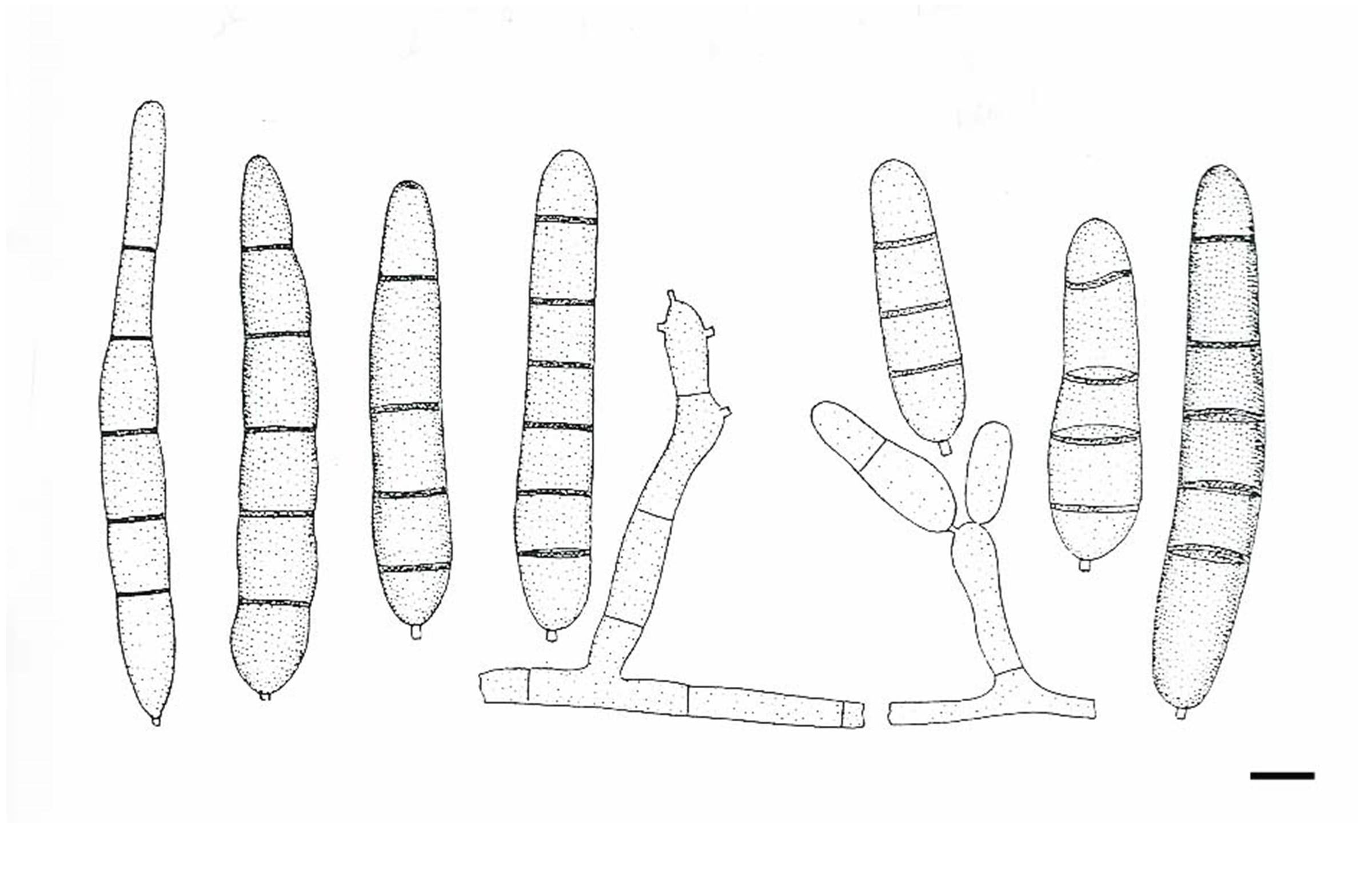
Saprobic or parasitic in marine habitats. Sexual morph: unknown. Asexual morph: Conidiophores 30–40 × 2–4 µm, simple or branched, septate or not, straight or flexuous, often nodose with conspicuous, brown pigmentation at the apical region; at times reduced to conidiogenous cells. Conidiogenous cells terminal or lateral, with denticles aggregated at apex, with prominent conidial scars, thickened but not darkened; sometimes proliferating with a new head or a short, inconspicuous sympodial rachis. Conidia 16–65 × 5–9 µm, produced holoblastically, on narrow denticle, smooth-walled, cylindrical to obclavate, straight or slightly flexuous, 1–7 transverse septa, pale to medium brown, often with dark septa (often constricted), and a darkened zone of pigmentation at the apex, and at the hilum, which is thickened, and somewhat protruding, with a minute marginal frill (Description of Scolecobasidium salinum from Ellis 1976).
Notes: Paradendryphiella was introduced by Woudenberg et al. (2013) to accommodate two species Dendryphiella arenariae and D. salina (= Cercospora salina). Paradendryphiella is characterised by simple or branched conidiophores with a new head or a short, inconspicuous sympodial rachis Conidiogenous cells and holoblastically produced cylindrical to obclavate, straight or slightly flexuous, 1–7 transverse septa, pale to medium brown conidia. The phylogenetic trees show that Paradendryphiella nested within the family Pleosporaceae and clustered sister to Pleospora senso stricto clade in Jones et al. (2008), Woudenberg et al. (2013) and as well as in this study. Therefore we accept Paradendryphiella as separate genus in the family Pleosporaceae.
Fig 1 Paradendryphiella salina (Redrawn from Scolecobasidium salinum in Ellis (1976), Fig 138c.). Scale bars: 5 μm