Obovoideisporodochium lithocarpi Z. X. Zhang, J. W. Xia & X. G. Zhang, sp. nov.
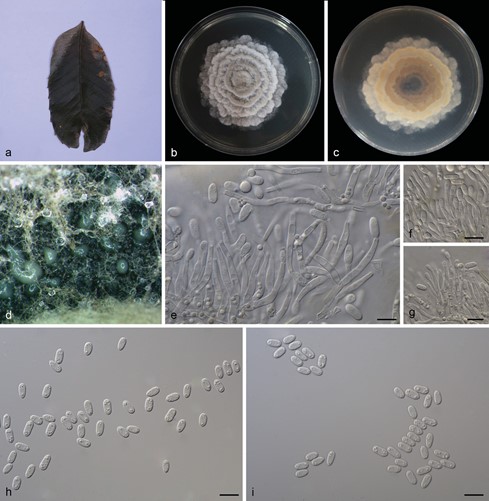
MycoBank number: MB 841104; Index Fungorum number: IF 841104; Facesoffungi number: FoF12723; Fig. 3
Type. China, Yunnan Province: Xishuangbanna Tropical Botanical Garden, Chinese Academy of Sciences, on diseased leaves of Lithocarpus fohaiensis (Fagaceae), 11 Sep 2020, Z. X. Zhang, (holotype HSAUP0748, ex-type living culture SAUCC 0748).
Etymology. Name refers to the genus of the host plant Lithocarpus fohaiensis.
Description. Asexual morph: mycelium consisting of septate, smooth and hyaline hyphae, thin-walled, 1.0–2.0 μm. Colonies on PDA incubated at 25°C in the dark with an average radial growth rate of 5–6 mm/d and reaching 75–80 mm diam. in 14 d, formed some conspicuous concentric circles, aerial mycelium cottony, white initial- ly, then becoming greyish-sepia. Conidiomata sporodochial, appeared within 20 days or longer, formed on agar surface, slimy, pale bluish-green, semi-submerged. Sporo- dochial conidiophores densely and irregularly branched, 12.0–26.5 × 1.5–3.0 μm, bearing apical whorls of 2–3 phialides; sporodochial phialides monophialidic, subulate to subcylindrical, 9.5–20.0 × 1.5–3.0 μm, smooth, thin-walled, tapering towards apex, swelling at base. Conidia formed singly, obovoid to ellipsoid, 5.5–8.0 × 2.5–4.0 μm, length/width ratio 1.7–3.1, hyaline, smooth, thin walled, apex obtuse, base with in- conspicuous to conspicuous hilum, 0.4–0.9 μm diam. Sexual morph: unknown.
Culture characteristics. Cultures incubated on MEA at 25°C in darkness, attaining 52.0–58.0 mm diam. after 14 d (growth rate 3.5–4.0 mm diam./d), grey- white to creamy white with irregular margin, spread like petals from the inside and outside, reverse dark to light brown, distributed in an irregular circle. Conidial formation not observed.
Additional specimen examined. China, Yunnan Province: Xishuangbanna Tropical Botanical Garden, Chinese Academy of Sciences, on diseased leaves of Lithocarpus fohaiensis (Fagaceae), 11 Sep 2020, Z. X. Zhang, HSAUP0745; living culture SAUCC 0745.
Notes. In the two phylogenetic trees (Figs. 1 and 2), Obovoideisporodochium lithocarpi is related to Racheliella wingfieldiana, Oblongisporothyrium castanopsidis, Paratubakia subglobosa and P. subglobosoides, but forms a separate single species lineage with full support (PP = 1, ML-BS = 100%). Furthermore, the conidia of O. lithocarpi (5.5–8.0 μm × 2.5–4.0 μm) are smaller than those of R. wingfieldiana (11.0–15.0 μm × 6.5–7.5 μm), Ob. castanopsidis (14.0–17.0 μm × 7.0–9.5 μm), P. subglobosa (10.0–13.0 μm × 8.0–11.0 μm) and P. subglobosoides (10.0–12.5 μm × 5.5–10.0 μm) and Racheliella, Oblongisporothyrium and Paratubakia spp. form crustose conidiomata and true pycnothyria.

Figure 1. Phylogram of Tubakiaceae, based on the concatenated ITS, LSU and rpb2 sequence alignment. The BI and ML bootstrap support values above 0.74 and 74% are shown at the first and second position, re- spectively. The tree is rooted to Greeneria uvicola (culture FI12007) and ex-type cultures are indicated in bold face. Strains from the current study are in red. Some branches were shortened for layout purposes – these are indicated by two diagonal lines with the number of times a branch was shortened indicated next to the lines.

Figure 2. Phylogram of Tubakiaceae, based on the concatenated ITS, tef1 and tub2 sequence alignment. The BI and ML bootstrap support values above 0.74 and 74% are shown at the first and second position, respectively. The tree is rooted to Greeneria uvicola (culture FI12007) and ex-type cultures are indicated in bold face. Strains from the current study are in red. Some branches were shortened for layout purposes
– these are indicated by two diagonal lines with the number of times a branch was shortened indicated next to the lines.

Figure 3. Obovoideisporodochium lithocarpi (SAUCC 0748). a infected leaf of Lithocarpus fohaiensis; b surface of colony after 15 days on MEA; c reverse of colony after 15 days on MEA; d conidiomata; e–g conidiophores, conidiogenous cells and conidia; h–i conidia. Scale bars: 10 μm (e–i).
