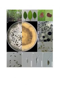
Neopestalotiopsis rhizophorae Norphanphoun, T.C. Wen & K.D. Hyde, Mycosphere 10(1): 545 (2020)
Index Fungorum number: IF 556437
Isolated from leaf spots of Rhizophora mucronata. Asexual morph: Conidiomata pycnidial, globose, brown, semi-immersed on PDA releasing black conidia in a slimy, globose, glistening mass. Conidiophores indistinct. Conidiogenous cells discrete to lageniform, hyaline, smooth and thin-walled, proliferating 1–2 times percurrently, collarette present and not flared. Conidia (19–)19.5–25(–26.5) × (5.6–)6–8(–9) μm (x̅ ± SD = 22.2 ± 1.9 × 7.2 ± 1.1 μm), fusiform to clavate, straight to slightly curved, 4-septate; basal cell obconic with a truncate base, hyaline or sometimes pale brown, thin- and smooth-walled, (3–)4–5(–5.7) μm long (x̅ ± SD = 4 ± 0.8 μm); three median cells (12.4–)13–15(–16.1) μm long (x̅ ± SD = 15 ± 1.1 μm), brown, septa and periclinal walls darker than rest of the cell, versicolored, wall rugose; second cell from base pale brown, (3.3–)4–5(–6) μm long (x̅ ± SD = 4.7 ± 0.7 μm); third cell brown, (3.3–)4–5(–6) μm long (x̅ ± SD = 4.8 ± 0.7 μm); fourth cell brown, (3.7–)4.5–5(–5.6) μm long (x̅ ± SD = 4.8 ± 0.6 μm); apical cell (2.8–)3–4(–5.2) μm long (x̅ ± SD = 4 ± 0.6 μm), hyaline, conic to acute; with 2–4 tubular appendages on apical cell, inserted at different loci but in a crest at the apex of the apical cell, unbranched, flexuous, (8.5–)11–29(–30) μm long (x̅ ± SD = 20 ± 6.9 μm); single basal appendage, tubular, unbranched, centric, (2.4–)3–11.5(–12) μm long (x̅ ± SD = 6.1 ± 2.8 μm).
Culture characteristics – Colonies on PDA reaching 0.5-2 cm diameter after 7 days at room temperature (±25 °C), colonies filamentous to circular, medium dense, aerial mycelium on surface flat or raised, with filiform margin (curled margin), fluffy to fairy fluffy, white from above and below; fruiting bodies black; reverse similar in color.
Material examined – Thailand, Kor Chang, Trat Province, leaf spots of Rhizophora mucronata, 27 April 2017, Norphanphoun C. KC23-1 (MFLU); living cultures, MFLUCC 17-1728; ibid. KC23-2 (MFLU); living cultures, MFLUCC 17-1729.
GenBank numbers – ITS: MN871770 (MFLUCC 17-1728), MN871771 (MFLUCC 17-1729); LSU: MN873006 (MFLUCC 17-1728), MN873007 (MFLUCC 17-1729); SSU: MN877440 (MFLUCC 17-1728), MN877441 (MFLUCC 17-1729); TEF1-α: MN871990 (MFLUCC 17-1728), MN871991 (MFLUCC 17-1729); β-TUB: MN871992 (MFLUCC 17-1728), MN871993 (MFLUCC 17-1729).
Known distribution (based on molecular data) – Thailand (Norphanphoun et al. 2019)
Known hosts (based on molecular data) – Rhizophora mucronata (Norphanphoun et al. 2019)
Notes – Based on multigene analyses and morphological characters, our isolate was identified as Neopestalotiopsis rhizophorae from leaf spots of Rhizophora mucronata in Thailand (Norphanphoun et al. 2019). Thus, these collections were considered as fresh collection of N. rhizophorae.

Fig. Neopestalotiopsis rhizophorae (MFLUCC 17-1728, MFLUCC 17-1729, fresh collections). a Collecting place. b, c Leaf spots of Rhizophora mucronata. d, e Culture on PDA (d-above, e-below). f–h Colony sporulating on PDA. i Conidiogenous cells giving rise to conidia. j–p Conidia. Scale bars: f = 200 µm, g = 1000 µm, h = 500 µm, i–p = 20 µm.
