Neopestalotiopsis musae Norphanphoun, T.C. Wen & K.D. Hyde, in Hyde et al., Fungal Diversity 80: 209 (2016)
Index Fungorum number: IF 552233; MycoBank number: MB 552233; Facesoffungi number: FoF 02363
Etymology – Named after the host genus on which the fungus occurs.
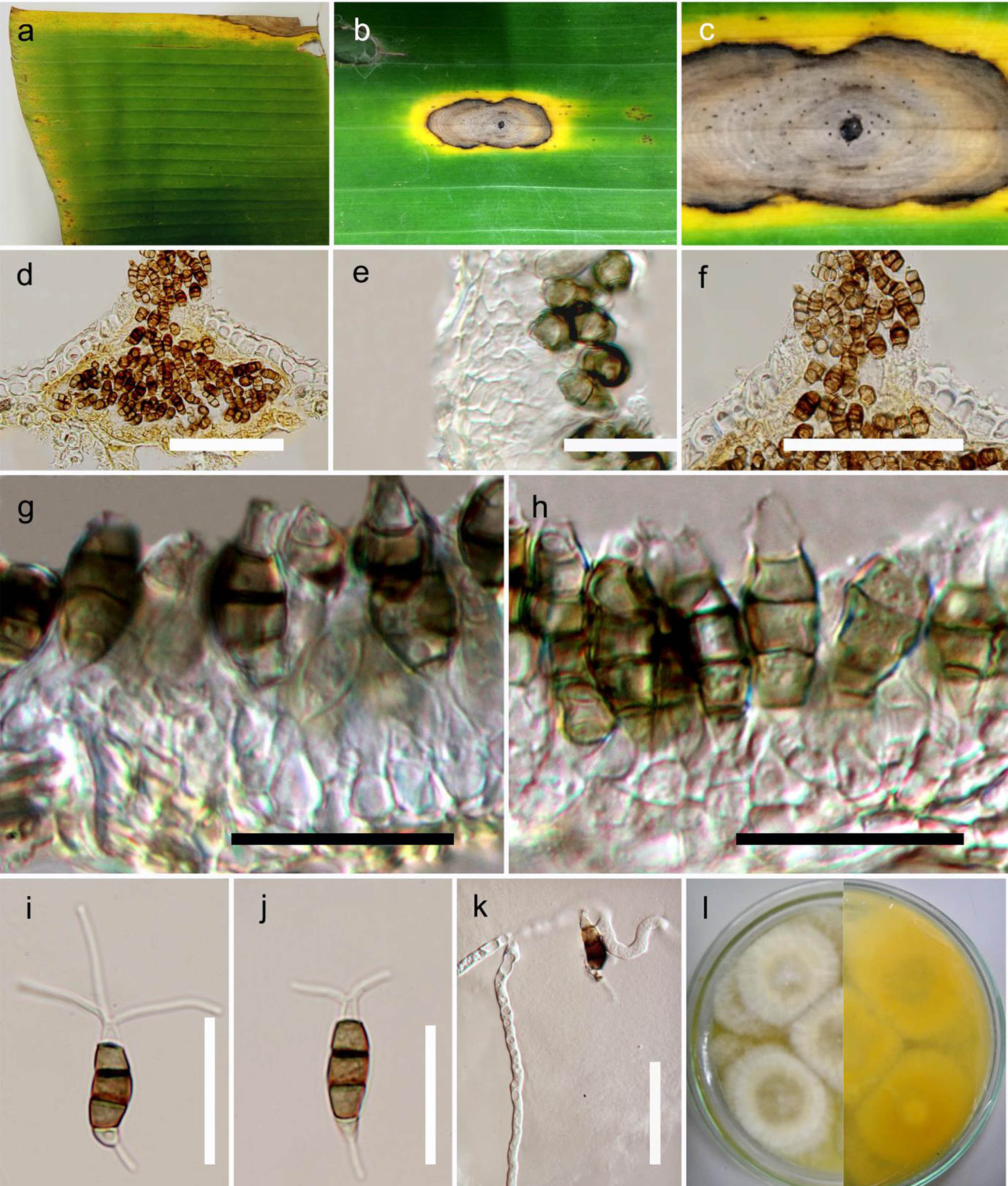
Pathogenic on Musa sp. Sexual morph Undetermined. Asexual morph Conidiomata 175–180 x 110–160 μm (x̄ = 175 x 140 μm ), acervuli, with basal stroma and lateral wall 1–3 cells thick; the wall cells hyaline, textura angularis. Conidiophores reduced to conidiogenous cells. Conidiogenous cells enteroblastic, discrete, simple, short, filiform. Conidia 18.6–25 x 4.1–5 μm (x̄ = 20.5 x 4.5 μm), fusiform to ellipsoid, straight to slightly curved, 4-septate; basal cell conic, hyaline, thin and smooth-walled, 3.2–5.2 μm long (x̄ = 3.5 μm); three median cells 12.1–16 μm long (x̄ = 15 μm), hyaline, versicolored, verruculose; second cell from base pale brown to olivaceous, 4.1–6.1 μm (x̄ = 5 μm); third cell pale brown to olivaceous, 3.2–6.1 μm (x̄ = 5.5 μm); fourth cell pale brown to olivaceous, 3.3–6 μm (x̄ = 4.5 μm); apical cell 3.6–5.2 μm long (x̄ = 3.7 μm), hyaline, cylindric to subcylindric; apical appendages 16.3–25 μm long (x̄ = 16 μm), 2–3 (mostly 3), basal appendage filiform, 4.6–10.3 μm long (x̄ = 5 μm).
Culture characteristics – Colonies on PDA, reaching 5 cm diam. after 10 days at 25 ºC, producing dense mycelium, circular, rough margin white, after 2 weeks, flat or effuse on the surface, without aerial mycelium.
Material examined – Thailand, Chiang Rai, Pha Chang, on leaf of Musa sp. (Musaceae), 4 February 2015, Chada Norphanphoun (MFLU 16-1279, holotype; KUN, isotype); ex-type-living cultures, MFLUCC 15-0776, KUMCC.
Notes – Molecular analysis provides good evidence that our strain belongs to Neopestalotiopsis and is closest to N. honoluluana Maharachch. et al. and N. zimbabwana Maharachch. et al. . However, N. honoluluana and N. zimbabwana has longer and wider conidia (x = 28 x 8.3 μm, = 25.3 x 7.7 μm, respectively) with N. honoluluana having three dark median cells, unlike N. musae which has smaller conidia (x = 20.5 x 4.5 μm), and pale-brown median cell of conidia (Maharachchikumbura et al. 2014b).
Fig. Neopestalotiopsis musae (holotype). a Leaf bright disease on banana. b, c Conidiomata on host substrate. d Cross section of conidioma. e Peridium. f Apex. g, h Conidiogenous cells with developing conidia. i–j Mature conidia. k Germinating spore. l Colony on PDA. Scale bars d, e = 50 μm, f–h = 20 μm, i–j = 15 μm, k = 25 μm.