Neoacanthostigma fusiforme Boonmee, Bhat & K.D. Hyde, sp. nov.,
MycoBank number: MB 550577; Index Fungorium number: IF 550577; Facesoffungi number: FoF 00187;
Etymology: in reference to the symmetrical fusiform ascospores.
Holotype: MFLU11–1146
Saprobic on dead wood. Sexual state: Ascomata (111-)126–138 µm high × 98–100(-125)µm diam. ( = 125 × 107 µm), superficial, solitary, scattered, globose to subglobose, reddish-brown to dark brown to black, surrounded by shining black setae (27-)48–73 µm long, tapering towards an acute tip, ostiolate. Peridium 13–14 µm wide, composed of several layers of brown cells of textura angularis. Hamatheciumcomprising ca. 1.5–2 µm wide, numerous,filiform, septate, branched, hyaline pseudoparaphyses. Asci 71–84 × 10–11(-12) µm (= 79 × 11 µm, n = 20), 8-spored, bitunicate, cylindric-clavate, with a short rounded pedicel, with thick and rounded apex, ocular chamber not observed. Ascospores (32-)40–48 × 3–4.5 µm ( = 43 × 4 µm, n = 20), fasciculate, cylindrical, narrowly fusiform, tapering towardsnarrow, subacute ends, straight to slightly curved, 5–7-septate, not constricted at septum, with mucilaginous pads at ends,hyaline, smooth-walled. Asexual state:hyphomycetous, helicosporous.Conidiophores up to 5 µm long, micronematous, holoblastic, polyblastic, dentate on creeping hyphae, hyaline, smooth-walled. Conidiahelicosporous, (17-)23–30 µm diam. when coiled, conidial filaments 2 µm wide, coiled in 1–2½ dimensional times, tapering toward rounded ends, multiseptate, not constricted at septa, hyaline, smooth-walled.
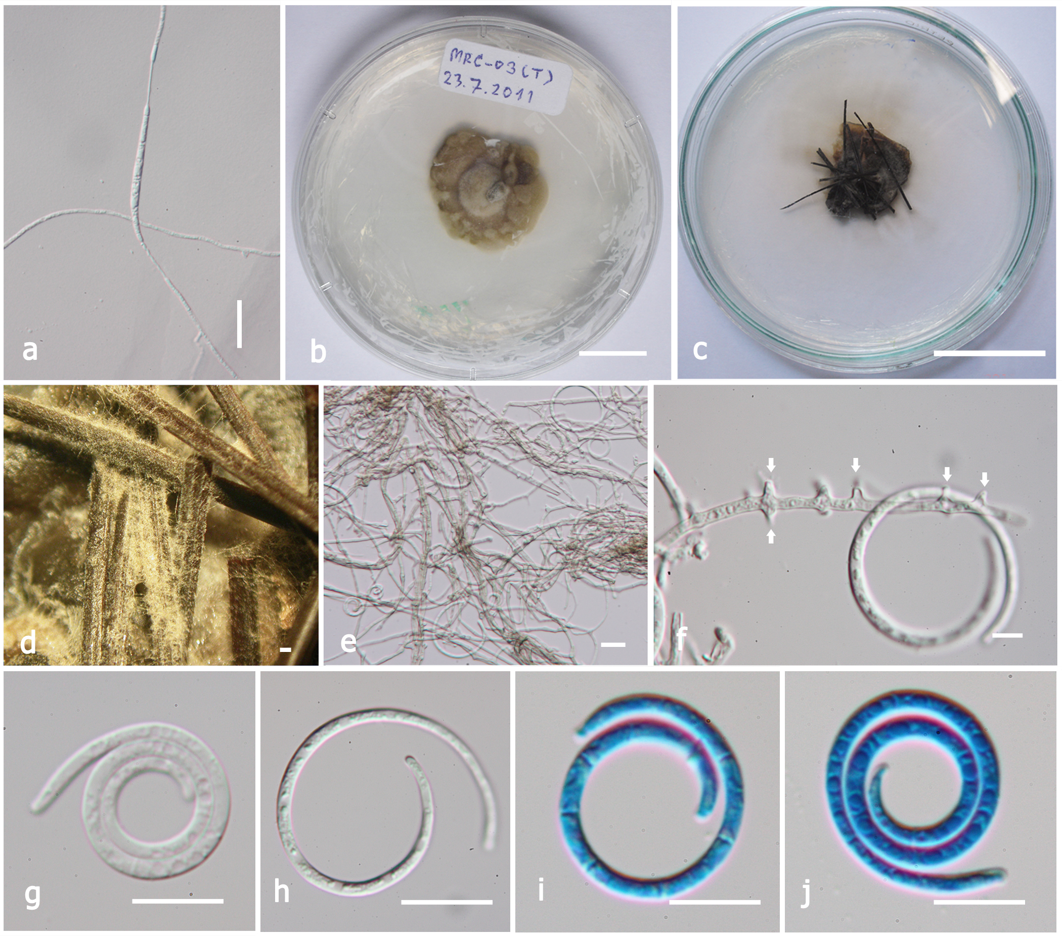
Cultural characteristics: Ascospores germinating on MEA within 8 h and germ tubes produced at both ends. Colonies growing on MEA slowly, less than 5 mm diam in 7 days at 28C, slightly raised-radially with lobate to entire edge, grayish to pale brown, laterally becoming dark brown. Mycelium developing on substrate, superficial, with hyaline to pale brown hyphae.
Fig. 1 Neocanthostigma fusiforme(MFLU11–1146, holotype). a Ascomata on substrate (arrows). b L.S. of ascoma. c Setae. d Peridium. e Pseudoparaphyses. f-h Asci. i-k Ascospores. Note spores becoming yellow when stained in Melzer’s reagent in Fig. k. Note the mucilaginous pads at the ends. Scale bars:a-b=100µm, c, f-h=50µm, d, i-k=20µm, e=5µm
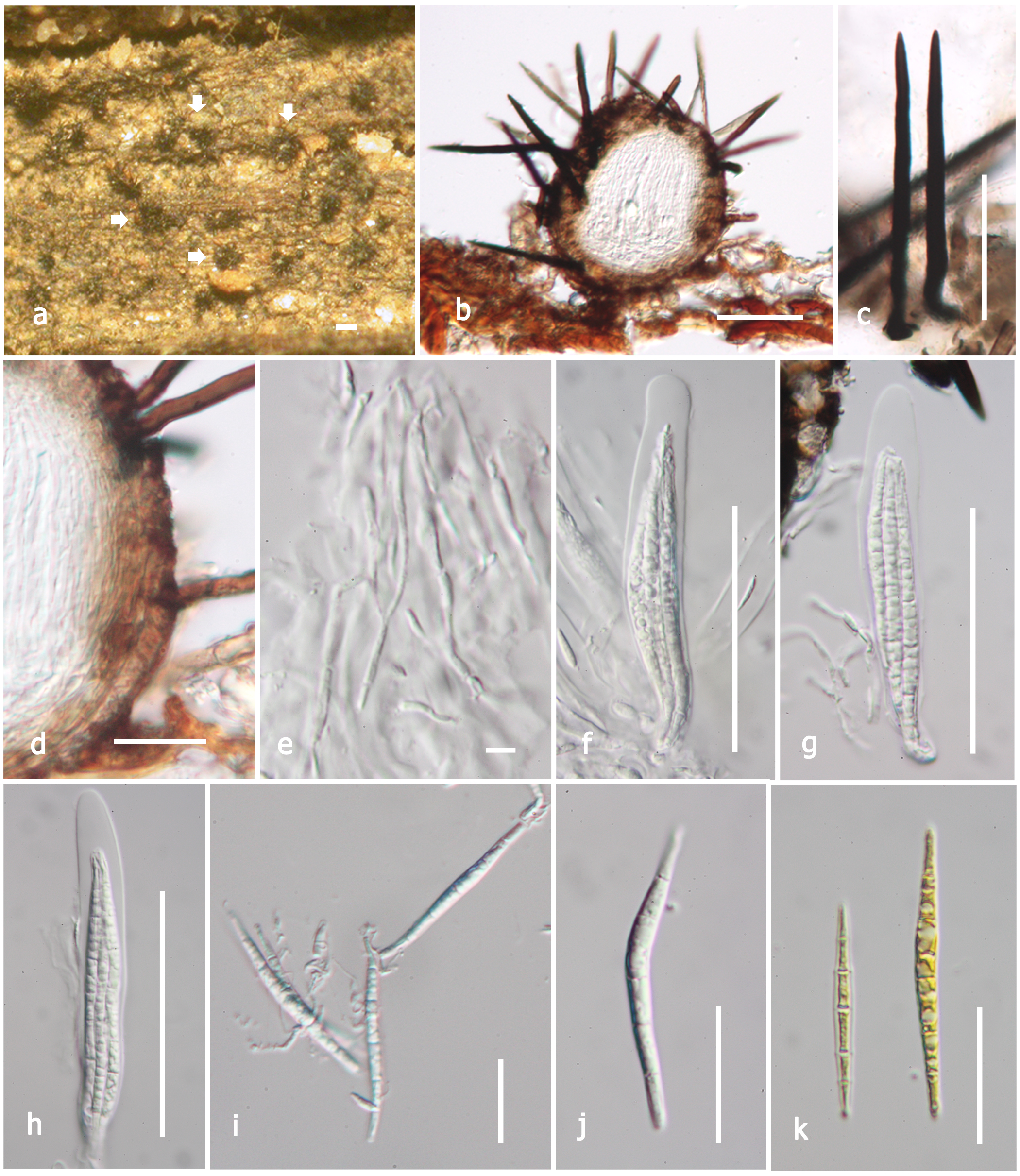
Fig. 2 Neocanthostigma fusiforme(MFLU 11–1146, holotype).a Germinating ascospore. b Colonies on MEA. Note colonies are grayish to pale brown. c, d Growth of asexual state on plant tissues produced on the media with and without substrate. e Aerial mycelium in culture.f Conidiophores formed on hyphae (arrows). g-j Conidia. Scale bars: a=20µm, b-d=10mm, e-f=5µm, g-j=10µm