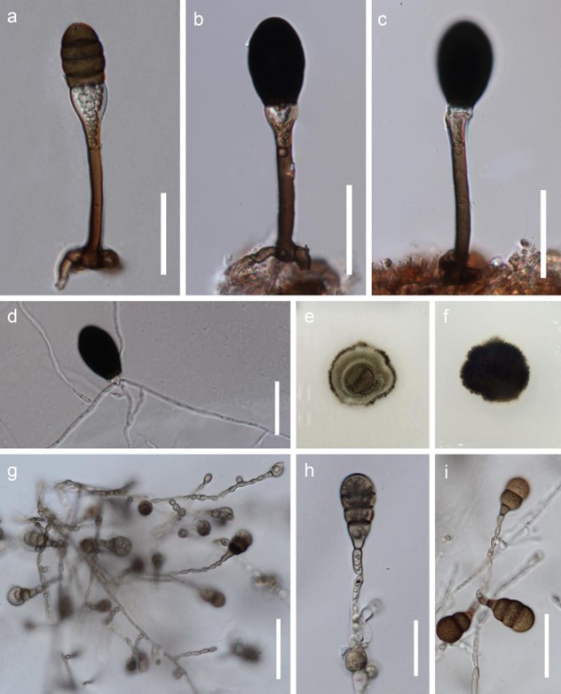
Mucispora infundibulata J. Yang & K.D. Hyde, sp. nov. Fig. 105
MycoBank number: MB 554769; Index Fungorum number: IF 554769; Facesoffungi number: FoF 04674;
Etymology – Referring to the infundibular conidiogenous cells.
Holotype – MFLU 18-0142.
Saprobic on decaying, submerged twigs. Sexual morph: Undetermined. Asexual morph: Colonies on substrate sparse, scattered, glistening, black. Mycelium mostly immersed, consisting of septate, smooth, pale brown to hyaline hyphae. Conidiophores macronematous, mononematous, solitary, straight, erect, smooth, mid brown, pale brown to hyaline and inflated at the apex, guttulated, 1–2-septate, 50–65 × 4–6 µm ( x = 60 × 5 µm, n = 10), 10–12.5 µm wide at the apex, with bulbous base. Conidiogenous cells monoblastic, integrated, terminal, cupulate or infundibulate, pale brown to hyaline, guttulate. Conidia acrogenous, smooth, broadly ellipsoidal to obovoid, 3-euseptate, constricted at the septa when young, with unobservable septa when mature, truncate at the base, (22–)29–34 × (15–)19–21 µm ( x = 31 × 20 µm, n = 10).
Culture characteristics – Conidia germinating on PDA within 24 h. Germ tubes produced from the conidial base. Colonies on PDA, reaching 5-10 mm diameter after one month at 25 °C in natural light, circular, with brown, greyish-green, white and dark brown mycelium from inner to outer circle; dark brown in reverse with entire margin. Hyphae subhyaline to pale brown, sometimes constricted at the septa, (4–)6–10(–14) µm wide. Conidiophores reduced to a monoblastic conidiogenous cell. Conidiogenous cells 8–10.5 × 3.3–4.8 µm, integrated, subhyaline to pale brown. Conidia (18–)23–35.5(–40) × (9.5–)12–17(–19) µm ( x = 31 × 14 µm, n = 20), pale brown to mid brown, 1–3-septate, mostly 2-septate, globose to obovoid, with cells becoming bigger towards the apical cell, smooth, constricted at the septa.
Material examined – THAILAND, Phang Nga Province, Bann Tom Thong Khang, on decaying wood submerged in a freshwater stream, 17 December 2015, J. Yang, Site 7-21-3, MFLU 18-0142, holotype, HKAS 102139, isotype; ex-type living cultures MFLUCC 16-0866, GZCC 17-0021.
GenBank numbers – ITS: MH457174, LSU: MH457139, SSU: MH457171.
Notes – Mucispora infundibulata resembles Melanocephala australiensis in conidial shape and size (Hughes 1979). Mucispora infundibulata was placed as a sister taxon to Mucispora phangngaensis with strong support (Fig. 10).Conidiophores of Melanocephala australiensis are initially 18–30 µm long, but can reach 100 µm by percurrent elongation, while conidiophores of Mucispora infundibulata are mostly around 60 µm long. The obvious difference is the inflated cupulate conidiogenous cells in Mucispora infundibulata compared to flared, percurrent proliferating in Melanocephala australiensis.

Figure 105 – Mucispora infundibulata (MFLU 18-0142, holotype). a-c Conidia and conidiophores. d Germinated conidium. e, f Cultures, e from above, f from below. g-i Asexual morph in culture. Scale bars: a-d, h = 30 µm, g, i = 50 µm.
