Montagnula acaciae Tennakoon, C.H. Kuo & K.D. Hyde, sp. nov.
MycoBank number: MB; Index Fungorum number: IF; Facesoffungi number: FoF 10801, FIGURE XX
Etymology: Name reflects the host genus Acacia.
Holotype: MFLU 18-2569
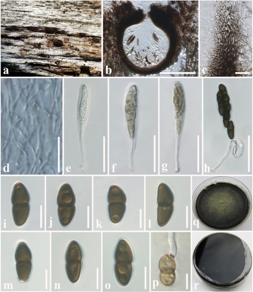
Saprobic on dead stem of Acacia auriculiformis A.Cunn. (Fabaceae). Sexual morph: Ascomata 140–180 μm high × 150–200 μm diam. ( = 160 × 175 µm, n = 10), solitary or scattered, coriaceous, immersed to semi-immersed, partly erumpent, visible as black dots on the host surface, unilocular, globose to obpyriform, brown to dark brown, ostiolate, with minute papilla, filled with hyaline cells. Ostiole papillate, protruding from substratum. Peridium 10–20 μm wide, outer layer comprising light brown to dark brown, thick-walled, cells of textura angularis, inner layer composed hyaline, flattened, thin-walled cells of textura angularis. Hamathecium comprising 1.5–2.5 μm wide, cylindrical to filiform, septate, branched, pseudoparaphyses. Asci 50–90 × 8–10 μm (= 72 × 9 μm, n = 20), 8-spored, bitunicate, fissitunicate, clavate to cylindrical, with a long pedicel, slightly curved, rounded at the apex and with a shallow ocular chamber. Ascospores 10–15 × 4–7 μm (= 12 × 5.8 μm, n = 30), overlapping, 1–2-seriate, hyaline or lightly pigmented when immature, fusiform, becoming light brown to dark brown when mature, fusiform to ellipsoid, 1-septate, guttulate, constricted at the septum, slightly wider upper cell and tapering towards ends, straight to slightly curved, without terminal appendages. Asexual morph: Undetermined.
Culture characteristics: Colonies on PDA, 15–20 mm diam. after three weeks at 25 °C, colonies from above: medium dense, circular, flat, surface smooth with entire edge, velvety, with smooth aspects, dark brown to black at the margin, grey in the centre; reverse: dark brown to black at the margin and centre, mycelium greenish to whitish grey.
Material examined: Thailand, Chiang Mai, Mushroom Research Center (MRC), on dead stem of Acacia auriculiformis (Fabaceae), 25 March 2016, D. S. Tennakoon, TD001A (MFLU 18-2569, holotype); ex-type living culture, MFLUCC 18-1636. ibid. 28 March 2016, TD001B (NCYU 19-0255, Paratype); ex-paratype living culture, NCYUCC 19-0087.
GenBank numbers: LSU: XXXX; SSU: XXXX; ITS: XXXX.; tef1-α: XXXX
Notes: The morphological characteristics of Montagnula acaciae tally with those described for Montagnula in having immersed to semi-immersed, brown to dark brown ascomata, clavate to cylindrical, long pedicellate asci with rounded at the apex and fusiform, light brown to dark brown, 1-septate ascospores (Ariyawansa et al. 2014b, Mapook et al. 2020, Hongsanan et al. 2020). Multi-gene phylogeny (LSU, SSU, ITS and tef1-α) generated herein indicates that Montagnula acaciae form an independent lineage sister to the clade formed by M. chromolaenicola, M. donacina, M. graminicola, M. puerensis, M. saikhuensis and M. thailandica with strong statistical support (96% ML, 1.00 BYPP, Fig. XX). Morphologically, Montagnula acaciae is different from M. saikhuensis and M. puerensis in having smaller ascomata (140–180 × 150–200 μm), whereas M. saikhuensis and M. puerensis have larger ascomata (400–450 × 400–500 μm and 300–600 × 230–380 μm). Montagnula donacina differs from M. acaciae in having carbonaceous ascostromata with flat bottom (Pitt et al. 2014). In addition, Montagnula graminicola can be distinguished from M. acaciae in having verrucous ascospores, not constricted in the middle and surrounded by obvious sheaths (Liu et al. 2015). Interestingly, we found that the morphological characteristics of Montagnula acaciae share similarities with M. thailandica, but they are phylogenetically distinct (Fig. XX). A comparison of the 550 nucleotides across the ITS (+5.8S) gene region of Montagnula acaciae and M. thailandica shows 12 base pair differences (2.18%). Furthermore, we compared Montagnula acaciae with M. thailandica based on base pair differences tef1-α gene region and there are 22 base pair differences (2.51%) across 880 nucleotides.

Fig. XX Montagnula acaciae (MFLU 18-2569, holotype). a Appearance of ascomata on substrate. b Vertical section through ascoma. c Peridium. d Pseudoparaphyses. e–h Immature and mature asci. i–o Ascospores. p A germinating ascospore. q Colonies from above (on PDA). r Colonies from below (on PDA). Scale bars: b = 100 µm, c = 8 µm, d–h = 30 µm, i–p = 5 µm.
