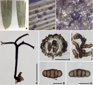
Meliola pseudosasae I. Hino, Bull. Faculty of Agriculture, Yamaguchi University 9: 882 (1958)
Index Fungorum number: IF 300776; MycoBank number: MB 300776; Facesoffungi number: FoF 02458
Epiphytes on the upper surface of leaves of Sasa borealis (Hack.) Makino & Shibata. Superficial hyphae branched, septate, darker at the septa, brown, with hyphopodia, hyphal setae present. Hyphopodia 10–11 μm diam. (x̅ = 10.5 μm , n = 20), head cell capitate, 22–25 μm long (x̅ = 23 μm, n = 20), alternate or opposite on hyphae, near to hyphal septum, 2-celled, upper cell cylindrical and curly, brown. Hyphal setae 170–210 μm high, 9–12 wide μm (x̅ = 180 × 10 μm, n = 10), arising from hyphae, comprising 2–3 arms, with 2–4-branches in each arm, acute and hyaline at the apex, septate, brown to dark brown, smooth-walled. Sexual morph Ascomata 97–135 μm high × 105–160 μm diam. (x̅ = 120 × 140 μm, n = 10), superficial on the surface of hosts, solitary to gregarious on superficial hyphae, globose to subglobose, thick-walled, ascomatal setae and appendages absent, surface of ascomata verrucose. Peridium 17–20 μm comprising dark brown cells of textura angularis when viewed in squash mounts, with two strata, outer stratum thick-walled, dark brown cells of irregular textura angularis, and inner stratum of flattened, hyaline cells. Hamathecium not observed. Asci not observed. Ascospores 45–52 μm high × 18–23 wide μm (x̅ = 50 × 22 μm, n = 10), 2-seriate, oblong to broadly cylindrical, hyaline to dark brown, 4-septate, constricted and darker at the septa, smooth-walled, ends rounded. Asexual morph Undetermined.
Material examined – CHINA, Xishuangbanna, on living leaves of Sasa borealis (Pinaceae), 22 November 2015, R. Phookamsak, Xb002 (MFLU 16-2136, in KIB, reference specimen designated here).
Notes – Our fresh specimen is similar to Meliola pseudosasae both in the host (Sasa borealis) and morphology. It has hyphal setae with 2–3 arms and with 2–4 branches at the apex, superficial hyphae with hyphopodia and 4-septate, brown ascospores as in the protologue. We were unable to isolate our specimen in culture, thus, DNA was extracted from ascomata and ascospores directly. Phylogenetic analysis indicates that our collection clusters with members of Meliolaceae within the subclass Meliolomycetidae, Sordariomycetes. Meliola pseudosasae is related to M. clerodendricola Henn. strain, which was published as a reference specimen by Hongsanan et al. (2015b). Asteridiella, Appendiculella and Endomeliola species clustered with Meliola species. Thus, further molecular data are needed to clarify if they are polyphyletic. We designate our collection as a reference specimen (sensu Ariyawansa et al. 2014b).

Fig. 1. Meliola pseudosasae (MFU 16-2136, reference specimen). a Herbarium specimen. b Appearance of ascomata on host leaf. c Ascoma.
d Setae. e Vertical section of ascoma. f Superficial hyphae. g, h Ascospores. Scale bars: d, e = 50 μm, f = 10 μm, g, h = 20 μm
