Magnibotryascoma kunmingense Mortimer, Frontiers in Microbiology: 9 (2021)
MycoBank number: MB 838522; Index Fungorum number: IF 838522; Faces of Fungi number: FoF 10662; Fig. **
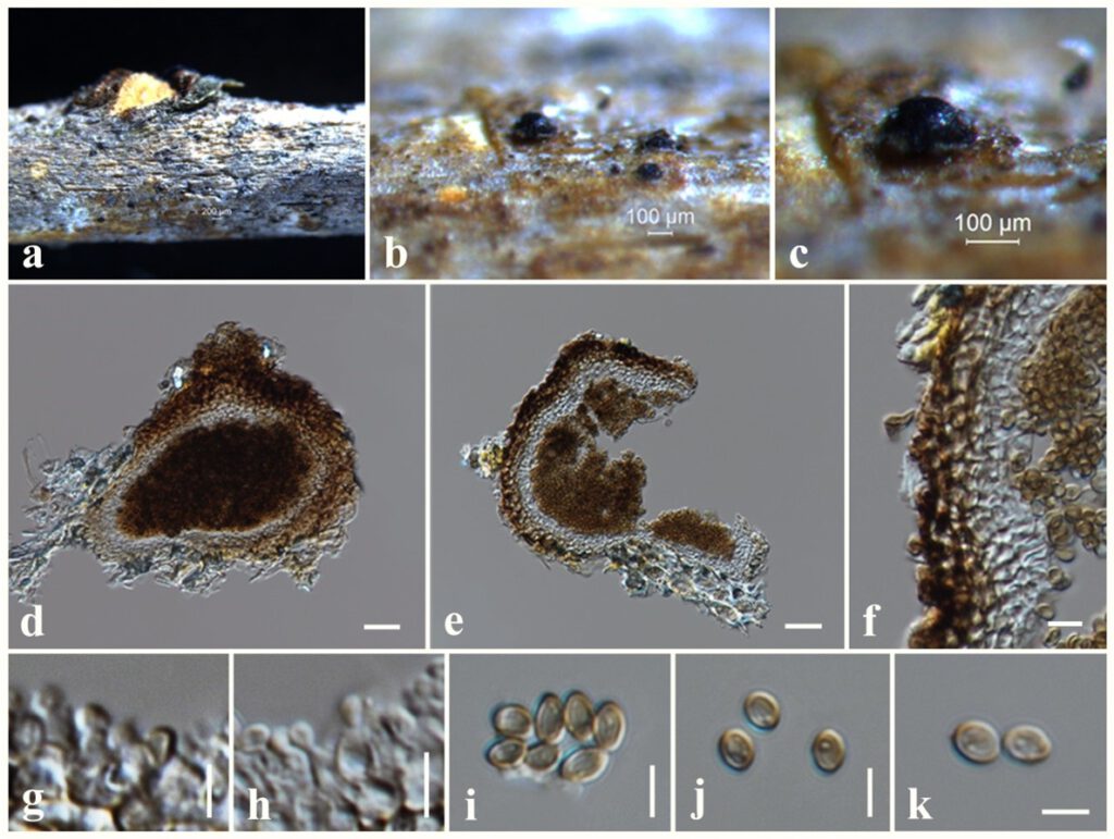
Saprobic on dead twigs attached to Magnolia sp. Sexual morph: Undetermined. Asexual morph: coelomycetous, Conidiomata 100–130 μm high × 115–140 μm diam. ( = 120 × 130 µm, n = 10), pycnidial, solitary, aggregated, uniloculate, semi-immersed to erumpent, globose to subglobose, coriaceous, dark brown to brown, papillate, with a central ostiole. Conidiomatal wall 15–23 μm wide, thick, 2-layered, with outer layer composed of light brown to brown cells of textura angularis, lined with a hyaline inner layer bearing conidiogenous cells. Conidiophores reduced to conidiogenous cells. Conidiogenous cells 3–5 × 3–4 μm ( = 4.5 × 3.5 μm, n = 20), enteroblastic, annelledic, discrete, cylindrical to oblong, hyaline, arising from the inner layer of pycnidium wall. Conidia 3–5 × 2.5–4 μm ( = 4.5 × 3 μm, n = 30), subglobose, oval, guttulate, hyaline when immature, pale brown at maturity, aseptate, smooth-walled.
Culture characteristics – Colonies on PDA reaching 20 mm diameter after 1 weeks at 25°C, colonies from above: cream, circular, flat, slightly raised, dense, white at the margin; reverse: pale brown from the centre of the colony, white at margin.
Material examined – THAILAND, Chiang Mai Province, dead twigs attached to Magnolia sp. (Magnoliaceae), 13 September 2017, N. I. de Silva, NI196 (MFLU 18-1295), living culture, MFLUCC 18-0708.
Known hosts and distribution – On dead twigs of Machilus yunnanensis and Acer cappadocicum in China (Mortimer et al. 2021), dead twigs (attached to the tree) of Magnolia sp. in Thailand (this study).
GenBank numbers – LSU: OL830817, ITS: OL966951, SSU: *******, tef1: *******, rpb2: ******.
Notes – The morphology of our collection (MFLU 18-1295) resembles Magnibotryascoma kunmingense in having semi-immersed to erumpent, globose to subglobose conidiomata, cylindrical to oblong, hyaline conidiogenous cells and subglobose, oval, pale brown, aseptate conidia (Mortimer et al. 2021). In phylogeny, our collection nested within other Magnibotryascoma kunmingense isolates in a well-supported clade (100% ML, 1.00 BYPP). Therefore, we introduce our collection as new host record of Magnibotryascoma kunmingense from Magnolia sp. in Thailand.

Figure * – Magnibotryascoma kunmingense (MFLU 18-1295). a Specimen. b, c Appearance of conidiomata on substrate. d, e Vertical sections through conidioma. f Peridium. g, h Conidiogenous cells. i–k Conidia. Scale bars: b, c = 100 μm, d, e = 20 μm, f–k = 5 μm.
