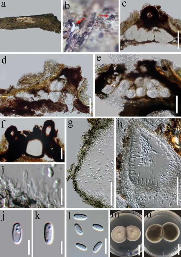
Loculosulcatispora thailandica G.C. Ren & K.D. Hyde, sp. nov. (Fig. 2)
MycoBank number: MB 557581;Index Fungorum number: IF 557581, Facesoffungi number: FoF 07978;
Etymology:—“thailandica” referring to the country where the species was first collected.
Type:—THAILAND. Chiang Rai Province: Mae Fah Luang University, on decaying woods, 13 May 2019, Guang-Cong Ren, RMFLU19006-1 (MFLU 20-0440, holotype; HKAS 107317, isotype), ex-type living cultures, MFLUCC 20-0108=KUMCC 20-0159.
Saprobic on woody litter. Sexual morph: undetermined. Asexual morph: coelomycetous. Conidiomata 200–300 × 44–57 ( x = 29.8 × 52.6, n = 4) μm, single or multiple, black, papillate, central. Conidiomata wall 10–40 μm thick, 2–5 layered, outer layer comprising brown to black cells of textura angularis, internal wall thin, comprising hyaline cells of textura angularis. Paraphyses 1–1.5 µm wide, sparse, aseptate, hyaline, unbranched, arising around inner wall of locules. Conidiophores reduced to conidiogenous cells. Conidiogenous cells 1.4–2.5 × 3.7–5.2 µm ( x = 1.8 × 4.4, n = 10), enteroblastic, phialidic, discrete, determinate, doliiform to cylindrical, hyaline, smooth-walled, arising from × 320–400 ( x = 235 × 360, n = 5) μm, conical or subglobose, black, scattered, semi-immersed to superficial, 3–9- loculate, ostiolate; each locule 40–50 × 48–58 ( x = 46.4 × 54.9, n = 10) μm, subglobose to ellipsoidal. ostioles 26–32 stratum. Conidia 5.7–8.5 × 2.2–3.1 ( x =6.9 × 2.6, n = 10) μm, 1-celled, oblong, hyaline, smooth-walled, guttulate.
Culture characteristics:—Colonies reaching 30 mm diam after 2 weeks at 25 °C, circular, white-grey at the center with white margin, floccose, umbonate, entire edge, white granular and circular crack on the surface, reverse atrovirens, darkening towards center and white at edge.
Notes:—The main morphological characters of Loculosulcatispora thailandica are multilocular conidiomata, conidiogenous cells arising from stratum, unbranched and aseptate paraphyses, 1-celled, oblong conidia, with 1–2 guttules. These features are morphologically different from others genera of Sulcatisporaceae (Crous et al. 2014, Tanaka et al. 2015).

FIGURE 2. Loculosulcatispora thailandica (MFLU 20-0440, holotype). a, b. Conidiomata on the natural wood surface (arrows). c–e. Sections through conidiomata. f. Ostiolar neck. g. Conidioma wall. h–i. Conidiogenous cells and developing conidia. j–l. Conidia. m-n. Culture characters on PDA. Scale bars: c–e=100 μm, f=50 μm, g=100 μm, h=50 μm, i=15 μm, j–k=5 μm, l=10 μm, m–n=20 mm.
