Lineostroma banksiae (Cooke) H.J. Swart, Trans. Br. Mycol. Soc. 91(3): 464 (1988), Index Fungorum number: IF 134625
≡ Didymosphaeria banksiae Cooke, Grevillea 19(no. 92): 90 (1891)
Pathogen on leaves of Banksia. ascomata in necrotic spots on upper surface of living leaves. Sexual morph: Ascostromata 75–125 µm high, 70–95 µm diam., linear, intraepidermal, at maturity breaking through the upper leaf surface, locules small, containing a few asci between paraphysoid threads, anastomosing, at maturity forming a small apical ostiole. Hamathecium of dense, septate, narrow, trabeculate pseudoparaphyses in a gelatinous matrix. Peridium 15–20 µm wide, inner layer hyaline, outer layer brown, comprising cells of textura angularis to textura globularis. Asci 45–70 × 8–10 µm (x̄ = 53 × 10 µm), 8-spored, bitunicate, cylindro-clavate, short pedicellate, apically rounded, with a minute ocular chamber. Ascospores 21–26 × 3–5 µm (x̄ =23 × 4 µm), uniseriate to biseriate, hyaline, becoming brown when mature, 1-septate, constricted at the septum. Asexual morph: Undetermined.
Material examined: AUSTRALIA. Victoria: on leaves of Banksia serrata (Proteaceae), 1892, Cooke (K(M) 143926, holotype).
Notes: The family Didymosphaeriaceae contains 13 sexual genera and two asexual genera (Ariyawansa et al. 2014a). Lineostroma banksiae has morphological characters that are distinct to other genera in the family, and hence we place it as a separate genus in the family Didymosphaeriaceae. Lineostroma banksiae is most similar to Phaeodothis in having linear, intra-epidermal ascostromata, trabeculate pseudoparaphyses, asci with a short pedicel and 1-septate ascospores. Phaeodothis tricuspidis differs in ascus arrangement and the peridium comprising hyaline, compressed cells (Ariyawansa et al. 2014a).
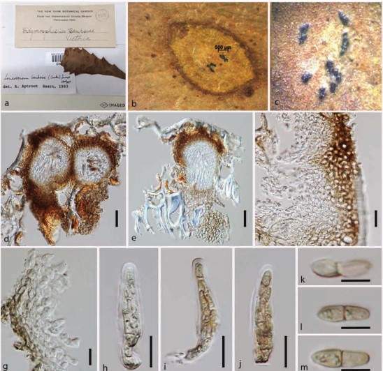
Fig. 2. Didymosphaeria banksiae (holotype). A Herbarium material. B Ascostromata on host surface. C Close up of ascostromata. D, E Section of the ascostromata. F Close up of the peridium. G Peridium. H–J Cylindrical to clavate asci with a small pedicel and ocular chamber. K–M Clavate-fusiform ascospores. Scale bars: D=100 µm, E=50 µm, F=5 µm, G=30 µm, I–L=10 µm.