Lasiosphaeris hispida (Tode) Clem., Gen. fung. (Minneapolis): [173] (1909)
Basionym: Sphaeria hispida Tode, Fung. mecklenb. sel. (Lüneburg) 2: 17 (1791)
Index Fungorum number: IF 531338, MycoBank number: MB 531338; Facesoffungi number: FoF 10021; Fig. 38
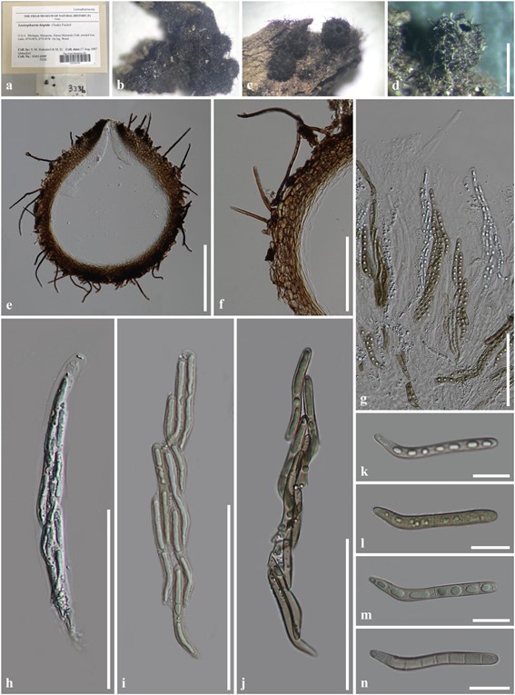
Saprobic on wood. Sexual morph: Ascomata 440 × 600 µm (x̄ = 520 µm, n = 10), perithecial, solitary or gregarious, superficial to semi-immersed, globose to sub-globose, black, membranaceous, ostiolate, with septate, brown, tapering hairs, 3.5–5.5 wide. Peridium 45–75 µm (x̄ = 62 µm, n = 30) wide, membranaceous, comprising two layers, outer layer composed of brown cells of textura angularis; inner layer composed of hyaline cells of textura prismatica. Paraphyses 2.5–5 µm wide, numerous, filiform. Asci (185–)210–230(–250) × 14–20 µm (x̄ = 220 × 17 µm, n = 30), 8-spored, unitunicate, cylindrical, pedicellate, apex rounded, with apical globule, apical ring distinct. Ascospores 60–70(–75) × 4.5–7.5 µm (x̄ = 65 × 6 µm, n = 50), bi-seriate, cylindrical to geniculate, slightly curved near the base, hyaline and aseptate when young, becoming pale brown and multi-septate when mature, with a large guttule in each cell, smooth-walled. Asexual morph: Undetermined.
Material examined – USA, Michigan, Marquette, Huron Mountain Club, around Ives Lake, 45º 0.00ʹ N, 87º 0.00ʹ W, on decayed wood, 17 August 1997, S.M. Huhndorf and M.H. Huhndorf (F-SMH3336).
Known hosts and distribution – On dead wood in Germany (type locality) and the USA (Fuckel 1870; Miller and Huhndorf 2005).
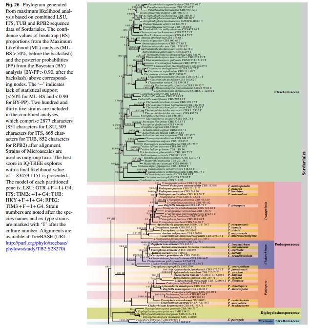
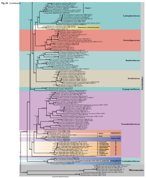
Notes – The molecular data of Lasiosphaeris hispida were provided (Miller and Huhndorf 2005; Vu et al. 2019). In this study, L. hispida is basal to L. arenicola and L. hirsute in Lasiosphaeridaceae (100%ML/1.00PP, Fig. 26). We could not obtain the type material. Therefore, we re-examined an authentic specimen collected by Huhndorf.


Fig. 38 Lasiosphaeris hispida (F-SMH 3336). a Herbarium material label. b Herbarium material. c, d Ascomata on host. e Ascoma in cross-section. f Peridium. g–j Asci. k–n Ascospores. Notes: Fig n soaked in Melzer’s reagent. Scale bars: d = 500 µm, e = 200 µm, f–j = 100 µm, k–n = 20 µm
