Lasiodiplodia fici Guiyan, X.; Manawasinghe, IS.; Phillips, AJL.; You, C.; Jayawardena, RS.; Hyde, KD.; Xiang, M.
MycoBank number: MB; Index Fungorum number: IF ; Facesoffungi number: FoF 10815;
Description: Epithet refers to the host genus from which the fungus was isolated
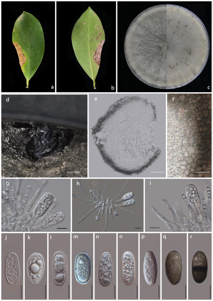
Pathogenic on Ficus altissima L. leaves. Sexual morph: Not observed. Asexual morph: Conidiomata pycnidial, produced on PDA within 4-8 wk, dark brown to black, unilocular, up to 3100 μmdiam, immersed in the needle tissue, globose to subglobose, ostiolate, wall composed of several layers of dark brown textura angularis. Peridium 20-50 µm, composed of thick-walled, brown-black cells of textura angularis, thin inner wall. Cylindrical, septate, unbranched, ends rounded, formed among conidiogenous cells. Conidiophores absent. Conidiogenous cells 20‒30 × 10‒15 µm (x̄ =27 × 12 µm,n=20), holoblastic, hyaline, smooth, thin-walled, cylindrical. Conidia 15–30 µm × 10–12 µm (x̄ =22 × 11 µm, n=150), initially hyaline, aseptate, ellipsoid to ovoid, thin-walled with granular content, rounded at apex, base round or truncate, becoming dark brown, 1-septate with longitudinal striations.
Culture characteristics: Colonies on PDA reach 7cm diameter after five days at 28°C. The top view filamentous, flat, and turbid. The aerial hyphae fluffy, dense and turn grey and black with time. The back becomes black.
Material examined: China, Guangdong Province, Guangzhou City, South China Botanical Garden on the dead leaves of Ficus altissima L. (Moraceae), May 12, 2021, X. Guiyan, (ZHKU 21-0092 Holotype), living cultures ZHKUCC 21-0125 ex-type; ZHKUCC 21-0126, ZHKUCC 21-0127.

Figure 3. Lasiodiplodia fici (ZHKU 21-0092; Holotype). a. Upper view of an infected leaf. b. Reverse view of an infected leave; c. Upper and reverse view of colonies on PDA after seven days. d. A pycnidia PDA after 28 days. e. Vertical section through a pycnidia. f. Pycnidial wall. g‒i. Conidia developing on conidiogenous cells. j‒r. Conidia. Scale bars: d= 1mm, e= 500um, f= 15um, g‒r= 10um.
