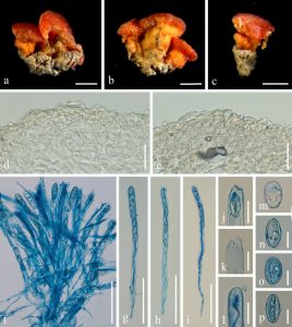
Kompsoscypha phyllogena (Seaver) Pfister, Mem. N. Y. bot. Gdn 49: 341 (1989)
Index Fungorum number: IF 136054
Saprobic on dead wood. Asexual morph: Undetermined. Sexual morph: Apothecia superficial, scattered to gregarious. Disc 0.5-1.5 mm high, 1-3 mm broad, orange to reddish, discoid to convex, hymenium orange, receptacle surface yellowish to orange. Stipe 200-500 µm long, 200-500 µm broad, yellowish. Medullary excipulum 100-150 µm broad, have cells of textura intricata, composed of 4-6 µm broad hyaline hyphae. Ectal excipulum 30-80 µm broad, composed of, 10-14 × 6-7 μm, hyaline cells of textura angularis. Stipitipellis 30-70 µm, comprised of 11-15 × 7-9 μm hyaline cells of textura angularis. Paraphyses filiform, 1-2 µm broad in the middle, hyaline, septate. Asci 267-315 × 12-15 µm, 8-spored, operculate, subcylindrical, J-. Ascospores [20/1/1, in H2O] (18.3-)19.2-21.5(-22.7) × (11.1-)11.7-13.2(-13.8) (Q = 1.41-2.00, Q = 1.65 ± 0.15), ellipsoid, equilateral uniseriate, inamyloid, guttulate, smooth-walled.
Material examined – China, Yunnan Province, Xishuangbanna, alt. 665 m, on unidentified dead wood, 9 June 2018, M. Zeng, Zeng 011 (HKAS 107663).
GenBank numbers – ITS: MT907443, LSU: MT907444.
Known distribution (based on molecular data) – China (this study), Puerto Rico (Romero et al. 2012)
Known hosts (based on molecular data) – dead wood or leaves (Romero et al. 2012, this study)
Notes – Kompsoscypha chudei is distinguished by orange to reddish apothecia, broad stipe, and ellipsoid, smooth ascospores with guttules. Ribes et al. (2015) discussed the differences between Kompsoscypha species. Based on the morphology and phylogeny, we introduced a new record of K. phyllogena from China.

Figure 1 . Kompsoscypha phyllogena (HKAS 107663, new geographical record). a–c Typical mature specimens. d Stipitipellis. e Receptacle surface of pileus. f Asci and paraphyses in Cotton blue. g–i Asci in Cotton blue. j–l Apices of asci in Cotton blue. m–p Ascospores in Cotton blue. Scale bars: a–c = 500 μm. d–e = 30 μm. f–i = 100 μm. j–l =20 μm, m–p =10 μm.
