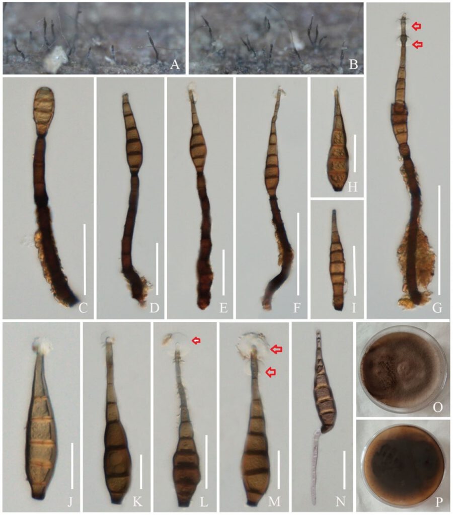
Kirschsteiniothelia xishuangbannaensis R.F. Xu, K.D. Hyde & Tibpromma, sp. nov. Fig 2.
MycoBank number: MB; Index Fungorum number: IF; Facesoffungi number: FoF 12758;
Holotype: ZHKU 22–0126
Etymology – The specific epithet “xishuangbannaensis” refers to Xishuangbanna, in Yunnan Province where the holotype was collected.
Saprobic on branch of rubber. Sexual morph: Undetermined. Asexual morph: Colonies effuse on natural substrate, solitary or scattered, hairy, brown to dark brown. Conidiophores 35–150 × 5–15 μm (x̅ = 104 × 10 μm, n = 10), brown to dark brown, septate, curved, solitary, wide and slightly swollen at base, tapering towards apex. Conidiogenous cells 10–50 × 8–10 μm (x̅ = 29 × 9 μm, n = 6), holoblastic, integrated, terminal, determinate, cylindrical or lageniform, smooth, brown to dark brown. Conidia 30–150 × 5–20 μm (x̅ = 93 × 16 μm, n = 20), acrogenous, solitary, yellow-brown to brown, subhyaline at apex, obclavate, rostrate, straight or curved, truncate at base, some have guttules, 3–8-septate, 1–2 hyaline globose/ampulliform mucilaginous sheaths around tip (mostly 1 mucilaginous sheath).
Culture characteristics – cultures on PDA, circular, flat, smooth, entire, reddish brown from above, the back is dark brown in the middle and the edges are reddish brown from below
Known distribution – widespread in tropical and subtropical regions.
Material examined – CHINA, Yunnan Province, Xishuangbanna, on branch of Hevea brasiliensis, 15 September 2021, Ruifang Xu, XSBNR–44, (ZHKUCC 22–0220, ex-type).
Notes – In the NCBI BLASTn searches of our new isolate (ZHKUCC 22–0220) show ITS sequence had 95.86% similarity and 35% query cover to K. submmersa (MFLUCC 150427: KU500570); LSU sequence had 98.23% similarity and 97% query cover to K. thailandica (MFLUCC 20-0116: MT984443) and SSU sequence had 99.80% similarity and 99% query cover to K. thailandica (MFLUCC 20-0116: MT984280). Our new strain K. xishuangbannaensis nested with the extant strains of K. thailandica in the phylogenetic analyses forming a clade with maximum statistical support (100% ML/1.00 PP). The sequence comparisons show 55 bp (12.11%, gaps 10 bp) differences in a 487 bp fragment of ITS, 13 bp (1.62%) differences in 803 bp fragment of LSU and 2 bp (0.19%) differences in a 1011 fragment of SSU between our strains and K. thailandica (MFLUCC 20-0116). Based on the guidance of Jeewon and Hyde (2016), a minimum of > 1.5% nucleotide differences in the ITS regions indicative of a new species. We also compared the morphology of our strain with K. thailandica and K. xishuangbannaensis has longer conidiophores than K. thailandica (35–150 μm vs. 55–93 μm), longer conidiogenous cells (13–50 μm vs. 9.5–21 μm), longer conidia (30–150 μm vs. 74–110 μm). While conidia of K. thailandica having olivaceous or brown with hyaline mucilaginous sheath around tip but K. xishuangbannaensis having yellow-brown to brown with 1–2 hyaline globose/ampulliform mucilaginous sheaths around tip. In addition, culture on PDA of K. xishuangbannaensis differ from K. thailandica (dark-olivaceous vs. reddish brown) (Sun et al. 2021).

Figs. 2 Kirschsteiniothelia xishuangbannaensis (ZHKU 22–0126, holotype) A, B Colonies on natural substrate. C–G Conidiophores with conidia. H–M Conidia (red arrows indicate mucilaginous sheath). N Germinated conidium. O, P Colony on PDA after one month. Scale bars: C, D, E, L = 50 μm, F, G = 100 μm, K, J, H, I, M, N = 30 μm.
