Juglanconis Voglmayr & Jaklitsch, Persoonia 38: 142 (2017)
MycoBank number: MB 819582; Index Fungorum number: IF 819582; Facesoffungi number: FoF 08844; 5 species with sequence data.
Type species – Juglanconis juglandina (Kunze) Voglmayr & Jaklitsch
Notes – Juglanconis was established by Voglmayr et al. (2017) to accommodate three combined species including J. juglandina (WU 35965, neotype), J. oblonga (MBT374386, lectotype), J. pterocaryae (TFM FPH2623, holotype) and a newly introduced species J. appendiculata (WU 35954, holotype). These species were previously placed in Melanconium but they are morphologically different from Melanconium sensu stricto in having irregular ornamentation on the inner surface of the conidial wall (Voglmayr et al. 2017). Fresh collections and updated morphological descriptions of Juglanconis juglandina and J. oblonga collected from Juglans regia in China were also provided by Fan et al. (2018). Subsequently, two new species Juglanconis japonica occurring on Pterocarya rhoifolia and J. pterocaryae occurring on Pterocarya fraxinifolia were introduced to this genus (Voglmayr et al. 2019b). To date, five species are accepted in Juglanconis and more fresh collections are needed (Voglmayr et al. 2019b). Currently Juglanconis species have calM, histone, ITS, LSU, ms204, rpb1, rpb2, tef1 and tub2 gene sequence data available for multilocus analyses (Voglmayr et al. 2019b).

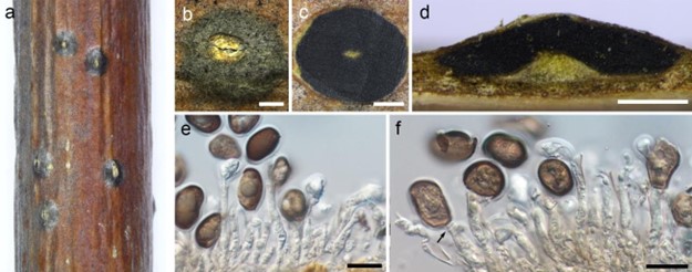
Figure 132 – Morphology of Juglanconis juglandina (Material examined – CHINA, Gansu Province, Qingyang City, Shishe village, on twigs and branches of Juglans regia, 14 July 2013, X.L. Fan, BJFC-S908). a, b Habit of acervuli on branches. c Transverse section through acervulus. d Longitudinal section through acervulus. e, f Conidiophores, conidiogenous cells and conidia. Annellides of conidiogenous cells are marked by black arrow in f. Scale bars: a-d = 0.5 mm, e, f = 20 μm.

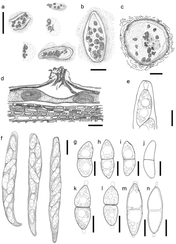
Figure 133 – Juglanconis juglandina (a-j), Juglanconis appendiculata (k-n) redrawn from Voglmayr et al. (2017). a, b Ectostromatic discs. c Transverse sections below ectostromatic disc. d Vertical section through pseudostroma, showing perithecia and entostroma. e Apical ring of asci. f Asci. g-n Ascospores. Scale bars: a = 1 mm, b, c = 0.5 mm, f =20 µm, e, g-n = 10 µm.

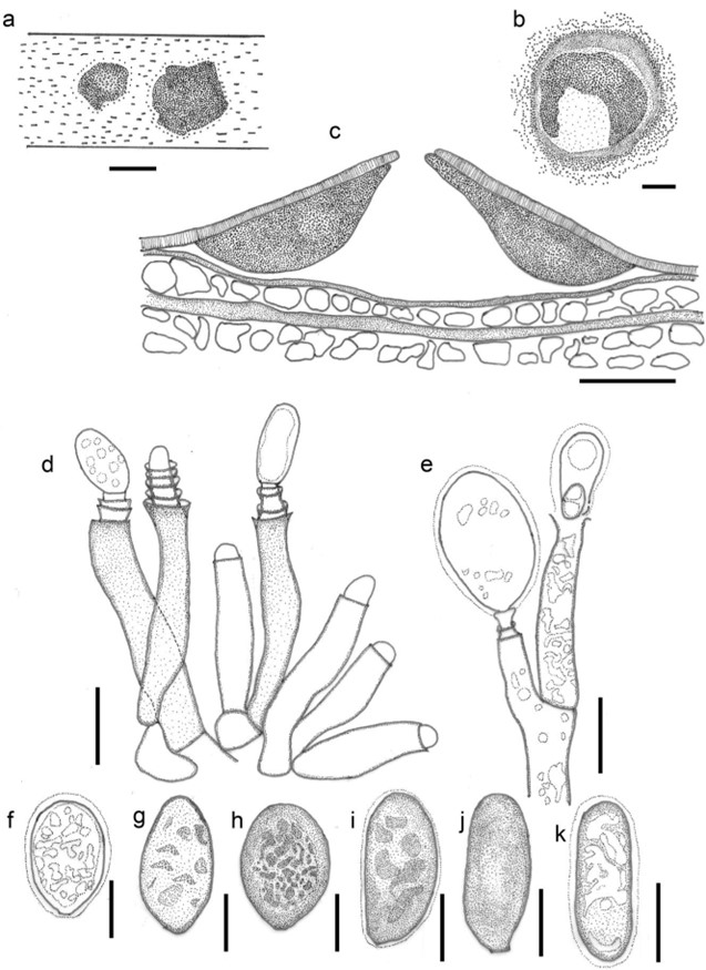
Figure 134 – Juglanconis juglandina (a-h), Juglanconis oblonga (i, j), Juglanconis pterocaryae (k) redrawn from Voglmayr et al. (2017). a Conidiomata on the host. b, c Transverse and vertical sections of conidiomata. d, e Conidiophore with conidia. f-k Conidia. Scale bars: a = 1 mm, b, c = 0.5 mm, d-k = 10 µm.
Species
Juglanconis juglandina
