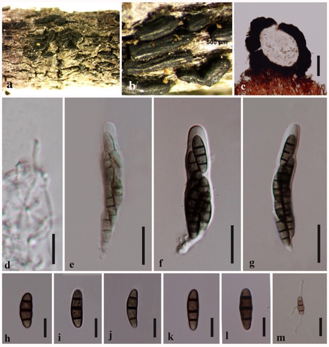
Hysterium angustatum Alb. & Schwein., Consp. fung. (Leipzig): 55 (1805). Fig. 39
MycoBank number: MB 221405; Index Fungorum number: IF 221405; Facesoffungi number: FoF 04579;
Saprobic on dead wood. Sexual morph: Hysterothecia 208–232 high × 256–284 wide × 500– 600 μm long (x̅ = 218 × 268 × 560 μm, n = 10), elongate and depressed conchate, scattered, superficial, base immersed in substrate, surface black, shiny, longitudinally striate, apex compressed, opening by a longitudinal slit. Peridium 40–60 μm (x̅ = 51, n = 15) carbonaceous, brittle, heavily pigmented, small prosenchymatous cells. Hamathecium comprising 0.5–1.5 μm, trabeculate, aseptate, branched, pseudoparaphyses, borne in a gelatinous matrix. Asci 55–70 × 8–12 μm (x̅ = 60 × 9 μm, n = 15), 8-spored, bitunicate, oblong to clavate, with a short narrow pedicel, apically thickened, with a distinct ocular chamber. Ascospores 15–19 × 4–6 μm (x̅ = 17× 5 μm, n = 25), crowded to 2–3-seriate, fusiform, hyaline when young and becoming brown at maturity, 3- septate, smooth-walled, ornamented, without mucilaginous sheath. Asexual morph: Undetermined.
Culture characteristics – Ascospores germinating on MEA within 24 hrs, slow growing at 18°C reaching 2 cm in 14 days, yellow at first, becoming ash when mature and reverse yellow.
Material examined – Australia, Melbourne, Mornington Peninsula, on dead wood, 10 March 2015, EBG Jones, GJ 107 (MFLU 16-2988; HKAS 96316)
Notes – We re-describe and illustrate Hysterium angustatum with a new strain. This is the first report of H. angustatum from Australia. Hysterium angustatum strains have little morphological variability in their spores, probably because of early speciation stages (Boehm et al. 2009a).

Figure 39 – Hysterium angustatum (MFLU 16-2988). a, b View of hysterothecia on host surface. c Section through hysterothecium. d Pseudoparaphyses. e−g Ascospores. h−l Asci. m Germinated ascospore. Scale bars: d = 10 µm, c = 100 µm, e−g, m = 20 µm, d, h−l = 10 µm
