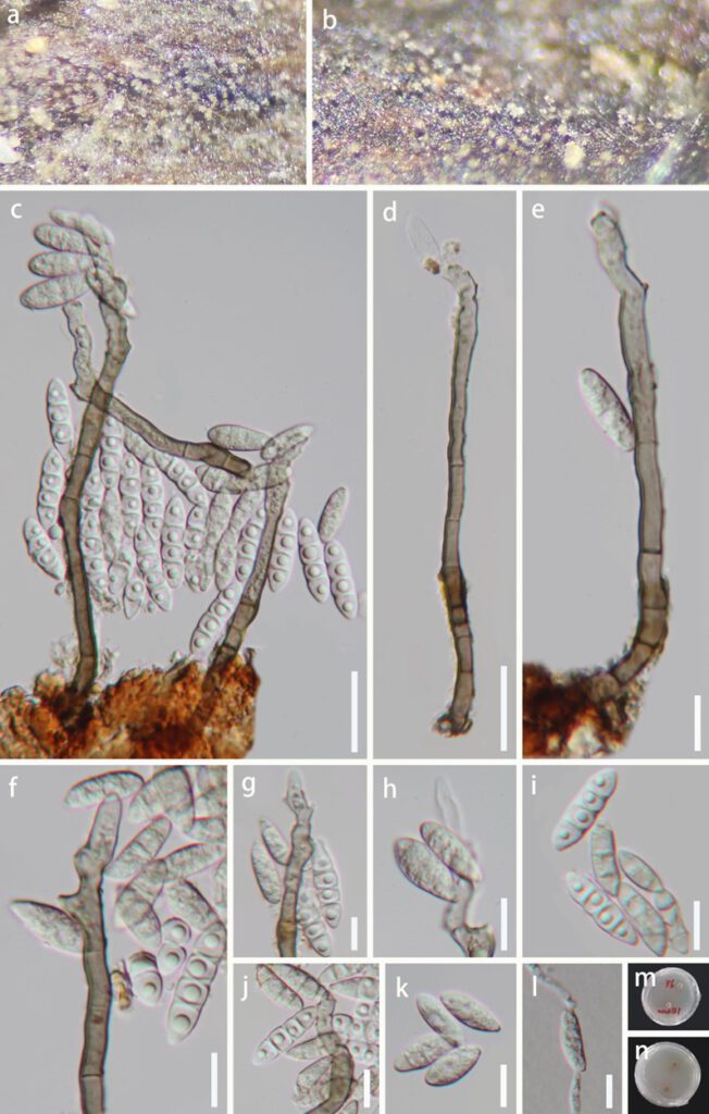
Hyaloguttulispora hainanensis J. Ma, K.D. Hyde & YZ. Lu., gen. nov. Fig. 2
MycoBank number: MB; Index Fungorum number: IF; Facesoffungi number: FoF 11002;
Etymology: The specific epithet ‘hainanensis’ refers to the locality where the fungus was collected, Hainan Province, China.
Holotype: HKAS 123764
Saprobic on decaying wood in freshwater habitats. Sexual morph: Undetermined. Asexual morph: Colonies on the natural substrate effuse, solitary, hairy, hyaline. Mycelium mostly superficial, consisting of branched, septate, smooth, pale brown to brown hyphae. Conidiophores 68.2–129.7 μm × 4.6–6.6 μm (x̅ = 90.9 × 5.6 μm, n = 30), macronematous, mononematous, solitary, erect, simple, straight or slightly flexuous, unbranched, smooth, cylindrical, 3–7-septate, thick-walled, smooth-walled, brown at the base, paler towards apex. Conidiogenous cells 25.2–62 μm × 3.7–5.9 μm (x̅ = 44.1 × 4.7 μm, n = 30), polyblastic, monoblastic, terminal, smooth walled, subcylindrical or tapering towards tip, pale-brown to subhyaline near base, hyaline toward apex, forming conidia sympodially on conspicuous denticles, bearing tiny, protuberant, circular scars. Conidia 16.6–27.8 μm × 4.2–6.2 μm (x̅ = 20.3 × 5.4 μm, n = 50) acrogenous, solitary, fusiform, clavate and suborbicular, 1–3-septate, hyaline, tapering and pointed at both ends, smooth‐walled, often with one guttule in each cell when young.
Culture characteristics: Conidia germinating on Potato Dextrose Agar (PDA) within 12 h and germ tubes produced from conidia. Colonies growing on PDA, reaching 8 mm diam. in 25 days at room temperature, circular, flat, lightly gray at the entire margin, brown in the center. In reverse, pale brown or brown.
Material examined: China, Hainan Province, Yanoda Tropical rainforest scenic area, on decaying wood submerged in a freshwater stream, 23 October 2021, Jian Ma, Y6, Y10-1 (HKAS 123764, holotype; GZAAS22-0077, isotype; HKAS 123763, paratype), ex-type living culture, GZCC22-0072.
GenBank accession numbers: (LSU), (ITS), (TEF1α) ON, (RPB2) ON.
Notes: Aquapteridospora is described herein as a monotypic genus that was collected on a submerged decaying wood in a freshwater stream in Hainan Province, China. The phylogenetic results confirmed that our collected two strains Hyaloguttulispora hainanensis formed a sister clade to the clade containing the genus Aquapteridospora (A. lignicola, A. bambusinum, A. aquatica and A. fusiformis) with good support (100% ML/1.0 PP, Fig. 1). Morphologically, H. hainanensis is similar to genus Aquapteridospora with macronematous, mononematous, solitary, erect, straight or slightly flexuous, unbranched, cylindrical, septate, smooth, thick-walled conidiophores, polyblastic, terminal, with several sympodial proliferations, bearing tiny, protuberant, circular scars conidiogenous cells and solitary, fusiform, acropleurogenous conidia. but differs by H. hainanensis has brown at the base, paler towards apex conidiophores, subcylindrical or tapering towards tip, pale-brown to subhyaline near base, hyaline toward apex conidiogenous cells, and clavate and suborbicular, septate, hyaline, tapering and pointed at both ends conidia[1]. Based on a pairwise nucleotide comparison of ITS, LSU, and TEF1α, H. hainanensis differs A. fusiformis in 73/ 582 bp (12.5%) for ITS, 21/ 823 bp (2.5%) for LSU and 62/ 860 bp (7.2%) for TEF1α, respectively. Therefore, we introduced Hyaloguttulispora as a novel genus based on the evidence of morphology and phylogeny.

Figure 2. Hyaloguttulispora hainanensis (HKAS 123764, holotype) a–b Colonies on natural substrate. c–e Conidiophores, conidiogenous cells bearing conidia. f–h, j Conidiogenous cells with attached conidia. j–k Conidia. l Germinating conidium. m, n Culture on PDA from above and reverse. Scale bars: e–l = 20 μm, c–d = 10 μm
