Hermatomyces thailandica Doilom D.J. Bhat & K.D. Hyde, sp. nov. Index Fungorum number: IF551966
Etymology: Name refers to the country in which the fungus was first collected.
Holotype: MFLU 15–3440
Saprobic on dead twigs of T. grandis. Sexual morph: Undetermined. Asexual morph: Conidia on natural substrate, superficial, gregarious, scattered, black. Mycelium 1.8–3.2μm wide, hyphae, superficial, branched, anastomosing, network, septate, reddish-brown. Conidiophores up to 30μm long, 2– 3.5 wide, micronematous to semi-macronematous, mononematous, straight or flexuous, slightly constricted at the septa, pale brown, gradually paler upwards, short, smooth, septate, unbranched, arising from hyphae, indeterminate. Conidiogenous cells monoblastic, integrated, terminal, cylindrical, hyaline to sub-hyaline, smooth. Conidia dimorphic, thick-walled, smooth, two types, mostly lenticular conidia; Lenticular conidia: (23–)28–29(−31) diam. × (14–)17–18(−20) μm thick in lateral views (x = 27 × 17μm, n = 50), muriform, blackish-brown at the central cells, subhyaline at peripheral cells, with peripheral cells surrounding central, dark brown in the center, pale brown next to the center, hyaline at the lower and upper cells, one column composed of 6–7 cells arranged in 2–4 rows, septate, constricted at the septa. Cylindrical conidia: (25–)30–31(−33) diam. × (16–)19–20(−21) wide in broadest part of lower cells μm, (x=30 × 19μm, n = 25), with 2 columns composed of 3 cells, with black peripheral cells, dark brown upper cells, usually with two cells hyaline in the lower cells, subglobose, swollen at the lower part, verrucose.
Culture characteristics: Conidia germinating on PDA with in 24 h. Germ tubes produced around conidia. Colonies on MEA reaching 21–23 mm diam. after 7 days in the dark at 25 °C (x = 21.5 mm, n = 5), edge entire, flat or effuse at the edge, convex with papillate surface at the center, medium dense, fluffy, aerial mycelium brownish grey (4E2) at the center, grey (4B1) at the edge from above; brownish grey (4F2) at the center and yellowish grey (4B2) at the edge from below.
Habitat: Known to inhabit dead moist twigs of T. grandis (current study).
Known distribution: Thailand (current study).
Material examined: THAILAND, Chiang Rai Province, Muang District, Mae Fah Luang University campus grounds, on dead moist twigs of T. grandis, 1 October 2014, M. Doilom, (MFLU 15–3440, holotype), ex-type living culture MFLUCC 14–1143, MKT 171/1, ICMP 21179, GenBank Accession No: ITS: KU144920, LSU: KU764692, RPB2: KU712488, SSU: KU712468, TEF1: KU872754; ibid. (MFLU 15–3441, paratype), ex-paratype living culture MFLUCC 14–1144, MKT 171/2, ICMP 21180, GenBank Accession No: ITS: KU144921, LSU: KU764693, RPB2: KU712489, SSU: KU712469, TEF1: KU872755; ibid. (MFLU 15–3442, paratype), ex-paratype living culture MFLUCC 14–1145, MKT 171/3, ICMP 21181, GenBank Accession No: ITS: KU144922, LSU: KU764694, RPB2: KU712490, SSU: KU712470, TEF1: KU872756.
Notes: Hermatomyces thailandica is introduced here as a second new species on T. grandis based on morphology, polymorphic nucleotide comparisons of the ITS, TEF1 and RPB2 sequence data (Table 3) and phylogenetic analysis (Fig. 5). Conidiophores of H. thailandica are shorter than those of H. tectonae, and the cylindrical conidia are also shorter and smaller, as well as lenticular conidia in lateral view (thick) are smaller. The colony colour and edge of H. thailandica can be differentiated from H. tectonae. Hermatomyces thailandica has an edge entire, is brownish grey (4E2) at the center, grey (4B1) at the edge from above, brownish grey (4F2) at the center and yellowish grey (4B2) at the edge from below but undulate to lobate and grey (1D1) above, pastel grey (1C1) from below after 7 days in the dark at 25 °C.
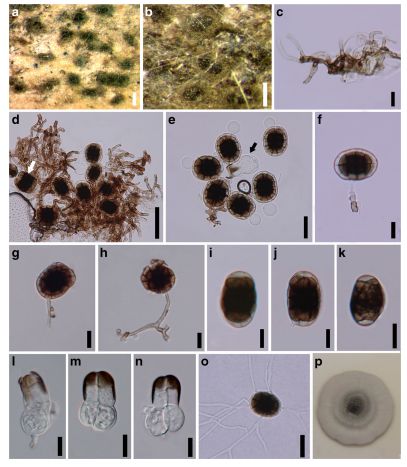
FIG Hermatomyces thailandica (MFLU 15–3440, holotype). a, b Conidia on host substrate. c Conidiophores with mycelia. d, e Lenticular conidia with lateral view (white arrow), cylindrical conidium (black arrow) with mycelia. f–h Lenticular conidia with conidiophores. i–k Lateral views of lenticular conidia. l–n Cylindrical conidia. o Germinated conidium. p Colony on MEA media after 7 days. Scale bars: a = 200 μm, b = 100 μm, c, f–n = 10 μm, d = 30 μm, o = 20 μm