Helicogermslita celastri (S.B. Kale & S.V.S. Kale) Lodha & D. Hawksw., in Hawksworth & Lodha, Trans. Br. mycol. Soc. 81(1): 91 (1983) (Figs. 66, 67)
Basionym: Amphisphaerella celastri S.B. Kale & S.V.S. Kale 1971
Saprobic on dead twigs. Sexual morph: Ascomata 0.4–0.6 mm diam., erumpent eventually becoming almost superficial, often persisting directly on wood, subglobose to pyriform, or mammiform, sometimes flattened at the base, shiny black, stromatic, aggregated into small groups (2–4), ostiolate, clypeate, dark brown to black, composed of dark hyphae and host cells. Ostioles 40–50 µm diam., scarcely protruding to distinctly short-papillate, visible as white spot. Peridium comprising 2–5 layers of thin-walled, dark brown to black, compressed, pseudoparenchymatous cells. Paraphyses 2.5–3 µm wide at the base, numerous, persistent, filiform, unbranched, septate. Asci 70–130 × 10–15 µm, 8-spored, unitunicate, elongate-cylindrical, short-pedicellate, lacks an apical ring. Ascospores 20–24 × 9–10 µm, uniseriate, broadly ellipsoidal, with rounded ends, fuscous brown to dark brown, or black, with helical or spiral germ slit, running 2–4 coils. Asexual morph: geniculosporiumlike.
Material examined – INDIA, Hyderabad, on unidentified wood, 6 Apr. 1981, C. Manoharachary (IMI 257189, isotype).
Notes – Holotype material of Helicogermslita celastri is housed at HCIO in India and this material is not available for loan. The other material at IMI was requested and studied here. Despite the fact that several collections of Helicogermslita celastri have been encountered from India no living culture is reported so far. Helicogermslita celastri has comparatively well-developed stromatic tissues than the other Helicogermslita species. Læssøe and Spooner (1994) have observed much broader ascospores than recorded by Hawksworth and Lodha (1983).
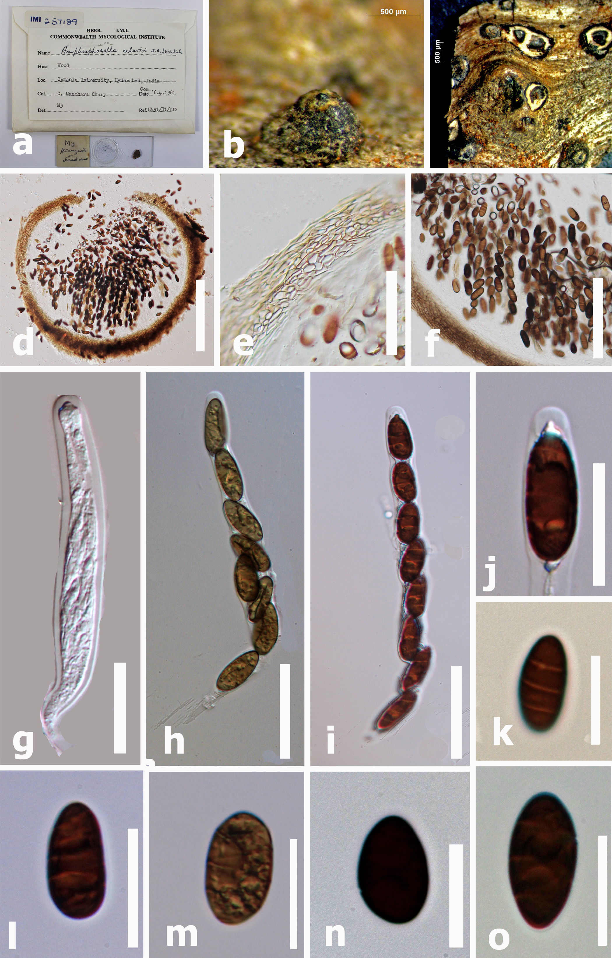
Fig. 1. Helicogermslita celastri (holotype) a. Herbarium details, b, c Stromata in wood, d. Cross section of stroma showing ascomata encased in stromal tissue. e Peridium. f Cluster of asci. g–i Asci, j Asci with apical ring bluing in Melzer’s reagent. k Ascospores showing the helical germ slit. l–o Ascospore. Scale bars d = 150 µm, c, d = 50 µm, g–h = 40 µm, j–o =10 µm.