Fuscostagonospora magnoliae N.I. de Silva, Lumyong & K.D. Hyde, sp. nov.
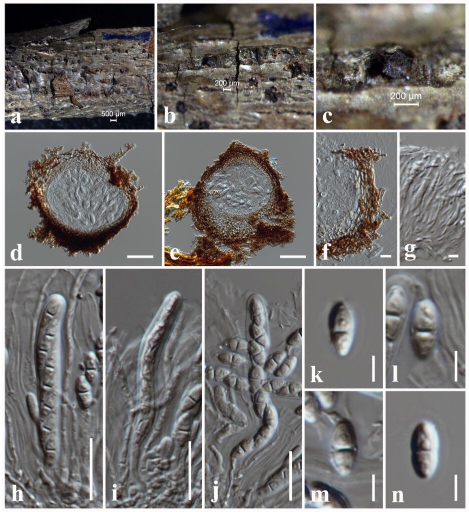
MycoBank number: MB 559516; Index Fungorum number: IF 559516; Facesoffungi number: FoF 10714; Fig. 6.13
Etymology: Name reflects the host genus Magnolia, from which the new species was isolated.
Holotype: MFLU 21-0218
Saprobic on dead twigs attached to Magnolia champaca. Sexual morph: Ascomata 160–190 μm high × 165–180 μm diam. ( = 180 × 170 µm, n = 10), solitary, scattered to clustered, semi-immersed to erumpent, black spots on host surface globose to subglobose, glabrous, uni-loculate, ostiole central with minute papilla. Peridium 25–30 μm thin-walled with equal thickness, composed of several layers of lightly pigmented to light brown to dark brown, textura angularis cells, cells towards the inside lighter, at the outside, darker and fusing with the host tissues. Hamathecium composed of dense, broad, 1–2 μm wide, filamentous, cellular pseudoparaphyses, with indistinct septa, not constricted at the septa, anastomosing at the apex, embedded in a hyaline gelatinous matrix. Asci 50–75 × 5–8 μm ( = 70 × 6 μm, n = 20), 8-spored, bitunicate, fissitunicate, cylindric-clavate, short pedicellate, with furcate to obtuse end, apically rounded with well-developed ocular chamber. Ascospores 9–12 × 4–6 μm ( = 10 × 5 μm, n = 40), overlapping, 1-seriate, ellipsoid to obovoid, hyaline, aseptate when young, becoming 1-septate, straight to slightly curved, smooth-walled. Asexual morph: Undetermined.
Culture characteristics – Colonies on PDA reaching 30 mm diameter after 1 weeks at 25°C, colonies from above: pale brown, circular, entire margin, slightly raised, dense at the centre, dark brown at the margin; reverse: brown from the centre of the colony, dark brown at margin.
Material examined – THAILAND, Chiang Rai Province, dead twigs attached to the host plant of Magnolia champaca (Magnoliaceae), 9 January 2019, N. I. de Silva, NI284 (MFLU 21-0218, holotype), ex-type living culture, MFLUCC 21-0176, NI285 living culture, MFLUCC 21-0177.
Notes – The morphological characteristics of Fuscostagonospora magnoliae resembles F. camporesii in having semi-immersed to erumpent, subglobose to globose ascomata, cylindric-clavate, short pedicellate asci and 1-septate, ellipsoid to obovoid ascospores (Hyde et al. 2020). However, Fuscostagonospora magnoliae can be distinguished from F. camporesii in having smaller asci (50–75 × 5–8 μm) and hyaline ascospores (9–12 × 4–6 μm), whereas F. camporesii has larger asci (80–90 × 8–9 μm) and light brown ascospores (13–15 × 6–6.5 μm) (Hyde et al. 2020). According to the multi-gene phylogenetic analyses of a combined LSU, SSU, ITS and TEF1-α sequence dataset, Fuscostagonospora magnoliae isolates nested sister to the F. camporesii with 83% ML and 0.99 BYPP support.

Figure 6.13 Fuscostagonospora magnoliae (MFLU 21-0218, holotype). a Specimen. b, c Appearance of ascomata on substrate. d, e Vertical sections through ascoma. f Peridium. g Pseudoparaphyses. h–j Asci. k–n Ascospores. Scale bars: a = 500 μm, b, c = 200 μm, d, e = 50 μm, f = 10 μm, g = 5 μm, h–j = 20 μm k–n = 5 μm.
