Fusarium brachygibbosum Padwick, Mycological Papers 12: 11 (1945)
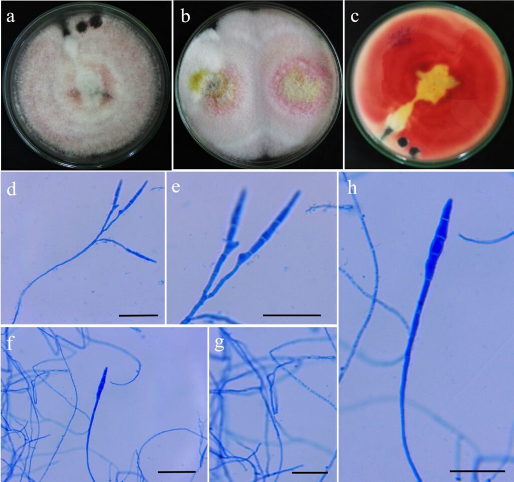
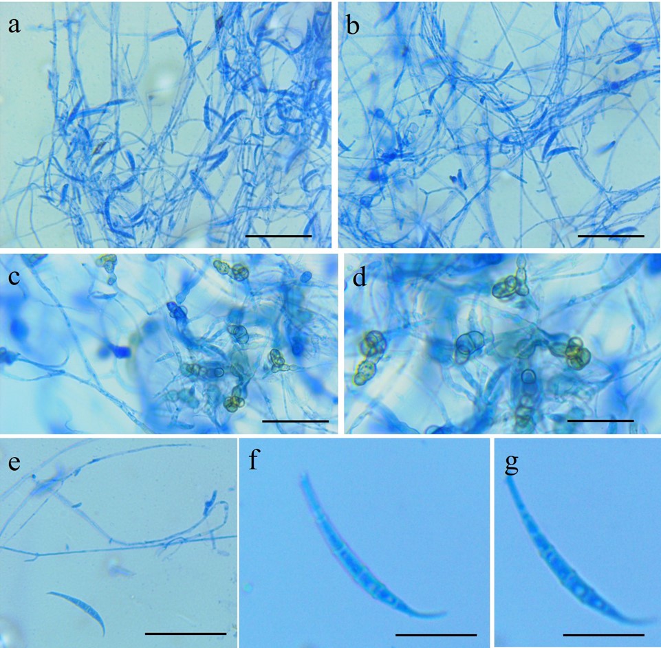
MycoBank number: MB 286508; Index Fungorum number: IF 286508; Faceoffungi number: FoF 11683 , Fig.1&2**
Pathogenic on roots of Vigna unguiculata. Sexual morph undetermined. Asexual morph Conidiophores 27–58 µm long, carried on aerial mycelium, unbranched or irregularly and/or sympodially branched bearing a terminal phialide. Conidiogenous cells 8–22×2–4 µm, polyphialide, subulate to subcylindical, smooth. Macroconidia 15.2–22 ×2-3 µm, hyaline, slightly curved with five distinct septa, wide central cells, slightly sharp apexes, basal cells with foot like shape. Microconidia rarely observed. Chlamydospores 6–24 µm diam. abundant, spherical o globose, smooth, slightly verrucose, formed terminally or intercalary in chains of two or three, wall 1–1.5 µm.
Cultural characteristics: Colonies on PDA reaching 90 mm at 28°C after 14 d in 12/12 dark, colonies appeared white to pink with abundant aerial mycelium.
Materials examined: India, Karnataka, Mysuru, Doddamaragowdanahally, diseased root of cowpea (Vigna unguiculata (L.) Walp.), S. Mahadevakumar, living culture (CPFb1)
GenBank Accession numbers: ITS: MT804589, MT804590, MT804591, MT804592; EF: OM938019, OM938020, OM938021, OM938022
Notes: F. brachygibbosum known to associated with 19 host plants of which two records are represented from India (Sorghum vulgare, Plasmopara viticola). This is the first record of F. brachygibbosum recorded on Cowpea (Fabaceae) from India (new host record).

Legend for Figure: Cultural and morphological features of Fusarium brachygibbosum: a – c pure cultures of F. brachygibbosum isolated on PDA medium (12 days old) (a-b Front view; c- reverse view); d – h Microscopic view of Fusarium brachyggibosum conidia structures observed under compound microscope (scale bar: d – h 20µm)

Legend for Figures: Morphological features of Fusarium brachygibbosum: a – b Conidial morphology of F. brachygibbosum under compound microscope; c – d hyphal structures and chlamydospores of F. brachygibbosum; e – f a single macroconidia enlarged (scale bar: a – d 50µm; e – 20µm; f – g 10µm)
