Distoseptispora thysanolaenae Goonas., Dayarathne, Phookamsak & K.D. Hyde, sp. nov.,
Index Fungorum number: IF555408
Etymology: Refers to the host from which the species was isolated
Holotype: KUN-HKAS 102247
Saprobic on dead culms of Thysanolaena maxima. Sexual morph: Undetermined. Asexual morph: Hyphomycetous. Colonies effuse, brown to dark brown, hairy or fluffy, arising from subiculum, with tufts. Mycelium
partly superficial, composed of branched, septate, smooth, brown hyphae. Conidiophores 30–80 × 3.5–5.5 μm (x̅ = 52 × 4.5 μm , n = 30), macronematous, mononematous, light to dark brown, 2–8-septate, smooth, usually flexuous or sometimes straight, unbranched, cylindrical, rounded at the apex. Conidiogenous cells monoblastic, integrated, terminal, determinate, hyaline to light brown, cylindrical. Conidia 21.5–80 × 6.5–12.8 μm (x̅ = 53 × 9.5 μm , n = 30), acrogenous, solitary, narrow and elongated obclavate, slightly curved, 8–14-distoseptate, thick-walled, light to dark brown, paler at the apex, tapering towards a rounded, flat apex, truncate with flat base, with conspicuous spore attachment loci, guttulate, smooth-walled.
Culture characteristics: Colonies on PDA reaching 43–45 mm diam. after 4 weeks at 20–25 ºC, colonies dense, circular, flat, surface slightly rough, rugose with edge entire, floccose to velvety; colony from above, greenish grey at the margin, brown-grey at the centre, from below dark grey at the margin, black at the centre; not producing pigmentation in PDA. Sporulation on PDA after two months. Conidiophores 10–30(–47) × 3–5 μm (x̅ = 21.4 × 4.6 μm, n = 25), macronematous, micronematous, mononematous, light to dark brown, 1–3-septate, smooth, usually flexuous or sometimes straight, unbranched, cylindrical, rounded at the apex. Conidiogenous cells monoblastic, integrated, terminal, determinate, hyaline to light brown, cylindrical. Conidia (20–)30–70(–243) × 5–8(–11) μm (x̅ = 53 × 8.4 μm, n = 30), acrogenous, varied in shape, elongate cylindrical to obclavate, lanceolate, rostrate, 6–22(–50)-distoseptate, light brown to dark brown, tapering towards a rounded, sometimes bulbous apex, truncate at the base, with conspicuous spore attachment loci, smooth-walled.
Material examined: CHINA, Yunnan Province, Xishuangbanna, Mengla County, Xishuangbanna Tropical Botanical Garden (XTBG), on dead culms of Thysanolaena maxima (Roxb.) Kuntze (Poaceae), 22 April 2017, R. Phookamsak, IS004 (KUN-HKAS 102247, holotype), extype living culture, KUMCC 18-0182 (IS004A), KUMCC 18-0183 (IS004B). GenBank numbers: ITS = MK045851, LSU = MK064091, TEF1-a = MK086031.
Notes: Distoseptispora thysanolaenae is the sixth species in this genus to be introduced from a terrestrial habitat. The previously introduced species are D. martini (J.L. Crane & Dumont) J.W. Xia & X.G. Zhang, D. tectonae Doilom & K.D. Hyde, D. tectonigena Doilom & K.D. Hyde, D. thailandica Tibpromma & K.D. Hyde and D. xishuangbannaensis Tibpromma & K.D. Hyde (Hyde et al. 2016; Xia et al. 2017; Luo et al. 2018; Tibpromma et al. 2018). Distoseptispora thysanolaenae can be distinguished from these species by its 2–8-septate conidiophore that are 30–80 × 3.5–5.5 μmand narrow elongated, obclavate, light to dark brown conidia that are 21.5–80 × 6.5–12.8 μm, 8–14-distoseptate and smooth-walled. Distoseptispora martini has longer conidiophores (50–110 × 3.5–4.5 μm), shorter and wider conidia (15–20 × 11–16 μm) that are ellipsoid, oblate or subglobose (Xia et al. 2017). Distoseptispora tectonae and D. tectonigena differ in their conidiophore dimensions (up to 40 × 4–6 μm, and up to 110 × 5–11 μm, respectively) and conidial dimensions ((90–)130–140(–170) × 13–14 μm, and 148–225(–360) × 11–12 μm, respectively) and number of septa. Distoseptispora thailandica and D. xishuangbannaensis have larger conidia (130–230 × 13.5–17 μm and 160–305 × 8–15 μm, respectively) and more septa (35–52-distoseptate, and up to 40-distoseptate, respectively) (Tibpromma et al. 2018). Phylogenetic analyses based on a combined ITS, LSU and TEF1-a sequence dataset show that our taxon clusters with other Distoseptispora species (Fig. 84). The species is sister to D. guttulata J. Yang & K.D. Hyde (MFLUCC 16-0183). A comparison of ITS and TEF1-a nucleotide bases shows that D. thysanolaenae differs from D. guttulata with 59 nucleotide bases of ITS and 64 nucleotide bases of TEF1-a. Distoseptispora thysanolaenae shares similar morphological features with D. guttulata but can be distinguished from the latter in having shorter conidia (21.5–80 μm versus 75–130 μm; Yang et al. 2018c). Therefore, following the guidelines of Jeewon and Hyde (2016) we introduce it as a new species.
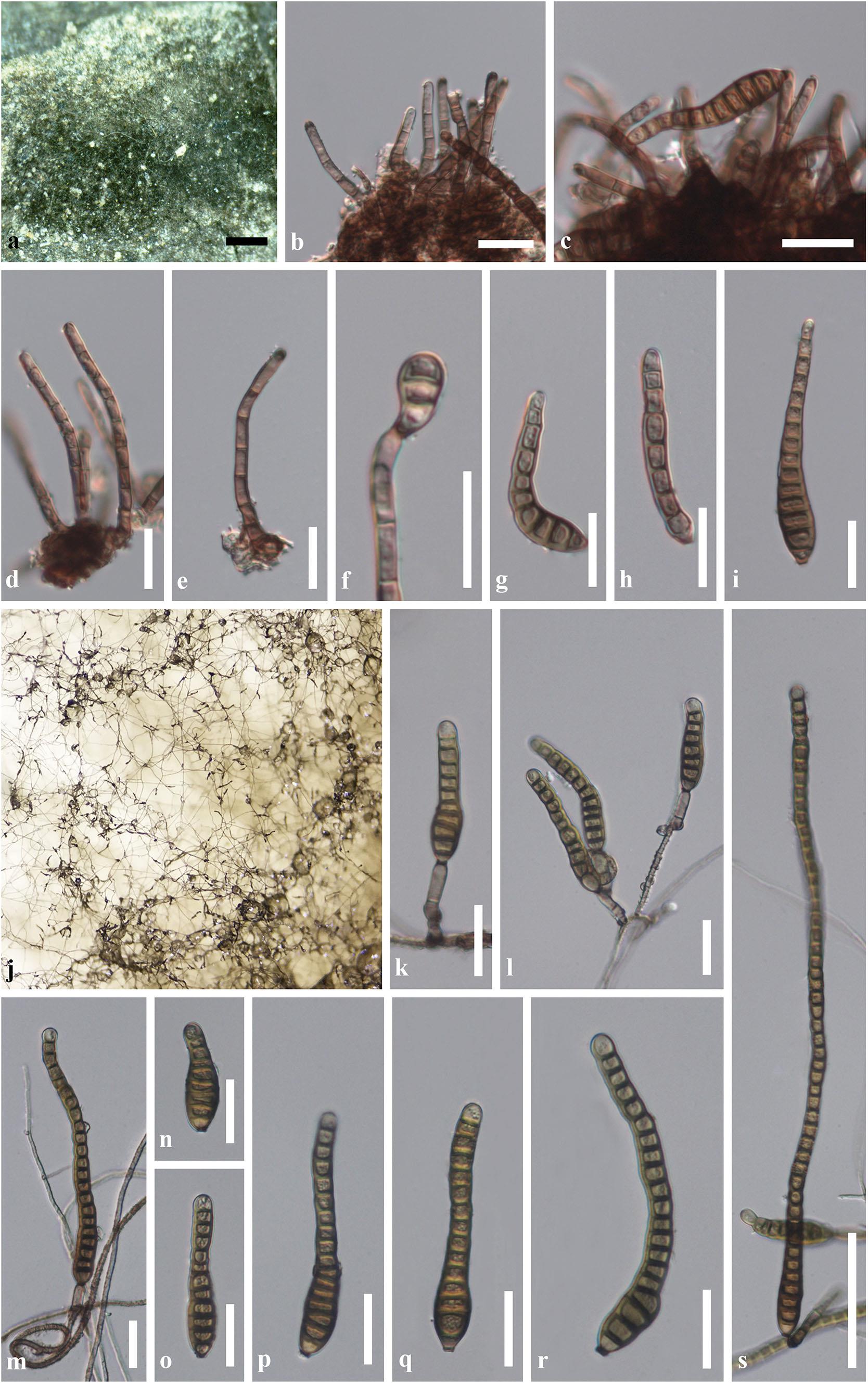
Fig. 85 Distoseptispora thysanolaenae (KUN-HKAS 102247, holotype). On host substrate: a Appearance of colonies on host surface. b, d, e Conidiophores. c, f Conidiophores with attached conidia. g–i Variable shapes of conidia. In vitro: j Sporulation of conidia on PDA after 4 weeks. k–mConidiophores with attached conidia. n–s Variable shapes of conidia. Scale bars: a = 500 μm, b–i, k–r = 20 μm, s = 50 μm.