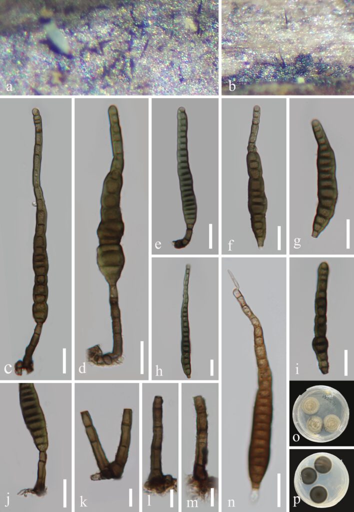
Distoseptispora wuzhishanensis J Ma, K. D. Hyde & Y.Z. Lu, sp. nov. (Figure 6).
MycoBank number: MB; Index Fungorum number: IF; Facesoffungi number: FoF 11336;
Etymology:‘wuzhishanensis’ named after the city from which the holotype was found.
Holotype: HKAS 123762
Saprobic on decaying wood submerged in freshwater habitats. Sexual morph: Undetermined. Asexual morph: hyphomycetous. Colonies on natural substrate superficial, effuse, solitary, smooth, septate, hairy, brown or dark brown. Mycelium mostly immersed, composed of branched, septate, smooth, pale brown to brown hyphae. Conidiophores 16.3–56.1 μm × 5.0–7.2 μm (x̅ = 35.24 × 6.2 μm, n = 25), macronematous, mononematous, cylindrical, erect, scattered or in small groups, straight or slightly flexuous, unbranched, Smooth, thick-walled, smooth-walled, brown at the base, pale brown towards the apex, 1-4-septate. Conidiogenous cells 6.6–10.7 × 4.4–6.2 μm (x̅ = 8.4 × 5.3 μm, n = 25), holoblastic, monoblastic, terminal, integrated, cylindrical, pale brown or brown, smooth. Conidia 76–142.9 × 10.9–17.1 μm (x̅ = 107.6 × 13.5 μm, n = 40), acrogenous, solitary, multi-distoseptate, obclavate, rostrate, straight or slightly curved, verruculose, guttulate, thick-walled, smooth-walled, pale brown or dark brown, olivaceous-green and yellow, usually up to 22 distoseptate, strongly constricted at septa, usually paler towards apex, rounded at apex, with a truncate base.
Culture characteristics: Conidia germinating on PDA within 12 h and germ tubes produced from conidia. Colonies growing on PDA, reaching 24 mm diam. in 25 days at 25°C, circular, flat, with dense, fluffy, olivaceous-green or brown mycelium on the surface; in reverse dark brown at the center, partly paler brown at the entire margin.
Material examined: China, Hainan Province, Wuzhishan National Nature Reserve, on decaying wood submerged in a freshwater stream, 26 December 2021, Xia Tang, W21 (HKAS 123762, holotype; GZAAS22-0082, isotype), ex-type living culture, GZCC22-0077.
GenBank accession numbers: (LSU), (ITS), (TEF1α) ON, (RPB2) ON.
Notes: In the phylogenetic analysis, Distoseptispora wuzhishanensis forms a related lineage with D. xishuangbannaensis, D. fasciculata, D.fluminicola and D. clematidis with moderate support (69% ML/1 BYPP). In a BLASTn search on NCBI GenBank, the closest match of the ITS and SSU sequence of our new isolate (D. wuzhishanensis) were 97.36% similarity across 97% of the query sequence and 98.69% similarity across 99% of the query sequence to D. sp. and D. fasciculata, respectively. The morphological characteristics of our new isolate match well with the generic concept of D. wuzhishanensis and is similar to D. xishuangbannaensis, D. fasciculata, D.fluminicola and D. clematidis in having mononematous conidiophore, monophialidic conidiogenous cells, and aseptate, acrogenous, solitary conidia. However, our new isolate differs from these new species by its obviously longer, scattered or in small groups conidiophores and multiple strongly constricted at septa conidia[8,9]. We, therefore introduced our new isolate as a novel species in the genus Distoseptispora based on morphological characters and phylogenetic evidence[10].

Figure 6. Distoseptispora wuzhishanensis (HKAS 123762, holotype) a–b Colonies on natural substrate. c–e Conidiophores and conidia. f–i Conidia. j–m Conidiophores and conidiogenous cells. n Germinating conidium. o, p Culture on PDA from above and reverse. Scale bars: c–j, n = 20 μm, k–m = 10 μm
