Distoseptispora rayongensis J. Yang & K.D. Hyde, sp. nov. Fig. 99
MycoBank number: MB 554770; Index Fungorum number: IF 554770; Facesoffungi number: FoF 04675;
Etymology – Referring to the collecting site in Rayong Province, Thailand.
Holotype – MFLU 18-1045.
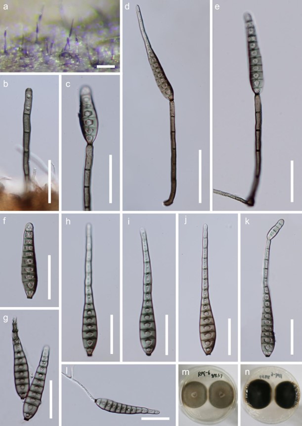
Saprobic on decaying twigs. Sexual morph: Undetermined. Asexual morph: Hyphomycetous. Colonies effuse, dark brown to black, hairy, glistening. Mycelium partly superficial, partly immersed in the substratum, composed of brown, septate, branched hyphae. Conidiophores macronematous, mononematous, solitary, cylindrical, straight or slightly flexuous, septate, brown, smooth, paler towards the apex, 75–125 × 3.5–5.5 µm (x̅ = 100 × 4.5 µm, n = 10), rounded at apex. Conidiogenous cells integrated, terminal, monoblastic, cylindrical, brown. Conidia acrogenous, solitary, obclavate or obspathulate, rostrate, mostly 9–13-euseptate, rarely 14–15-septate, pale brown or pale olivaceous, becoming paler or hyaline towards the apex, guttulate, smooth, (36–)60– 106(–120) × 9–14.5 µm (x̅ = 80 × 11.5 µm, n = 20), with a darkened scar at the base, sometimes with percurrent proliferation and forming another conidium from the conidial apex.
Culture characteristics – Conidia germinating on PDA within 24 h. Germ tubes produced from the conidial base. Colonies on PDA slow growing, reaching 5–10 mm diam. after one month at 25 °C in natural light, circular, aerial mycelium dense, brown; reverse dark brown, margin entire.
Material examined – THAILAND, Rayong Province, Klaeng, 12°78′N, 101°66′E, on decaying wood submerged in a freshwater stream, 24 April 2017, Y.Z. Lu, RAY-6, MFLU 18- 1045, holotype; ibid. HKAS102138, isotype; ex-type living cultures MFLUCC 18-0415; ibid. RAY-8, MFLU 18-1046, paratype; ex-paratype living culture MFLUCC 18-0417.
GenBank numbers – LSU: MH457137, SSU: MH457169, ITS: MH457172, tef1: MH463253, rpb2: MH463255 (MFLUCC 18-0415); LSU: MH457138, SSU: MH457170, ITS: MH457173, tef1: MH463254, rpb2: MH463256 (MFLUCC 18-0417).
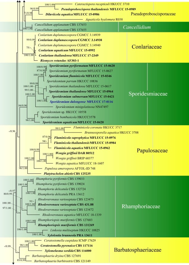
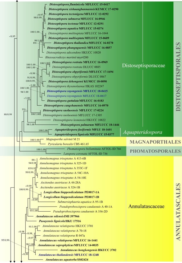
Notes – Distoseptispora rayongensis clustered within Distoseptispora with high statistical support in a sister clade to D. guttulata (Fig. 3). Distoseptispora rayongensis resembles D. guttulata and D. suoluoensis in having conspicuous long conidiophores and euseptate conidia of similar size. However, D. rayongensis can be distinguished from D. guttulata and D. suoluoensis in conidial colour and shape. Percurrently elongating conidia are observed in D. rayongensis and D. suoluoensis.

Figure 99 – Distoseptispora rayongensis (MFLU 18-1045 holotype). a Colonies on the woody substrate. b Conidiophore. c Conidiogenous cell with conidium. d, e Conidia and conidiophores. f-k Conidia. l Germinated conidium. m, n Cultures, m from above, n from below. Scale bars: a = 100 µm, b, c, e-l = 30 µm, d = 50 µm.

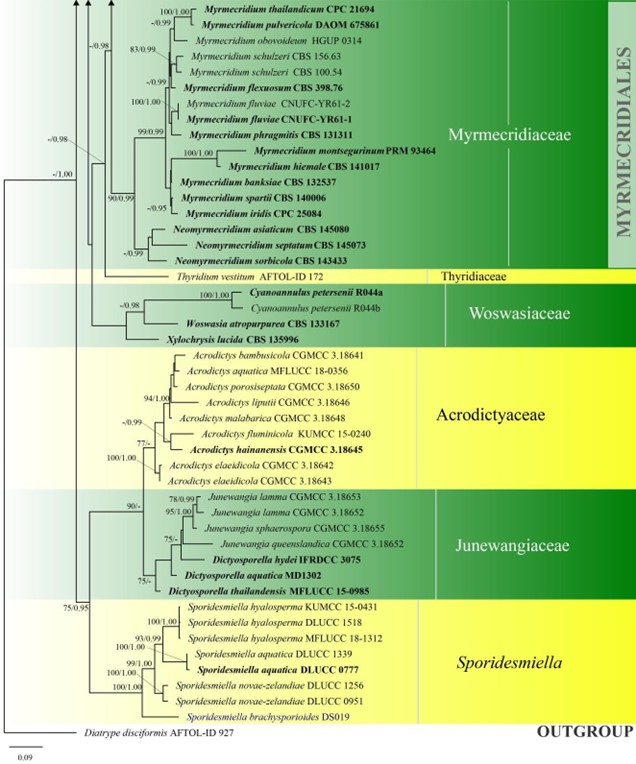
Figure 3 – Phylogram generated from maximum likelihood analysis based on combined LSU, SSU, ITS and rpb2 sequence data of Diaporthomycetidae. One hundred and ninety-three strains are included in the combined analyses which comprised 3545 characters (859 characters for LSU, 972 characters for SSU, 659 characters for ITS) after alignment. Single gene analyses were carried out and the topology of each tree had clade stability. Tree topology of the maximum likelihood analysis is similar to the Bayesian analysis. The best RaxML tree with a final likelihood value of – 68207.368884 is presented. Estimated base frequencies were as follows: A = 0.248206, C = 0.241993, G = 0.285500, T = 0.224301; substitution rates AC = 1.369088, AG = 2.887040, AT = 1.413053, CG = 1.152137, CT = 6.303994, GT = 1.000000; gamma distribution shape parameter a = 0.315782. Bootstrap support values for ML greater than 75% and Bayesian posterior probabilities greater than 0.95 are given near the nodes. The tree is rooted with Diatrype disciformis (AFTOL-ID 927). Ex-type strains are in bold. The newly generated sequences are indicated in blue.

Figure3 – Continued.

Figure3 – Continued.

Figure3 – Continued.
