Dichotomopilus ramosissimus (X. Wei Wang & L. Cai) X. Wei Wang & Samson, Stud. Mycol. 84: 217 (2016)
Index Fungorum number: IF 818869; Facesoffungi number: FoF 07245
Saprobic on Dolichandrone spathacea (L.f.) Seem. Asexual morph: Undetermined. Sexual morph: Ascomata 860 × 540 μm diam., superficial, subglobose or ovate, greenish, olivaceous to olivaceous grey in reflected light, ostiolate. Peridium wall composed of brown, angular or elongate hypha-like cells of textura angularis mixed with textura intricata. Terminal hairs dark brown, irregular in length, 3.2–6.5 μm diam. near the base, dichotomously branched at wide to acute angles starting near the bases, verrucose. Thinner hairs similar to the terminal ones but longer or seta-like, aseptate, 350 μm length. Lateral hairs are shorter spines. Asci not found. Ascospores 5.1–6.9 × 3–4.7 μm (x̅ = 6.0 × 4.0, n = 25), brown, ovate to broadly ovate, smooth-walled.
Culture characteristics – Colonies on malt extract agar olivaceous with an entire edge, about 53 mm diam. after 4 days of incubation at 28 °C, pale yellow to pale luteus, sparse and floccose aerial hyphae, without coloured exudates; reverse honey colored.
Material examined – India, Andaman and Nicobar Islands, North Andaman, Diglipur, Near Sita Nagar (13˚11’14.2” N 92˚53’11” E), isolated on decaying twig of Dolichandrone spathacea, 17 May 2018, M. Niranjan & V.V. Sarma, PUFNI 18725 (AMH-10059); living culture, NFCC-4521.
GenBank Accession numbers – ITS: MK990280, LSU: MK981539.
Known distribution (based on molecular data) – India, China, UK (https://www.ncbi.nlm.nih.gov/nuccore)
Known hosts (based on molecular data) – Clematis vitalba, Dolichandrone spathacea, Panax notoginseng (https://www.ncbi.nlm.nih.gov/nuccore).
Notes – Chaetomium ramosissimum was introduced by Wang et al. (2014) and later transferred to Dichotomopilus as D. ramosissimus (Wang et al. 2016). Dichotomopilus ramosissimus is characterised by its superficial, sphaerical or ovate ascomata and peridial wall consisting of textura intricata or epidermoidea cell layers. Our collection shows similar morphology for ascomata and also forms a yellow colony on malt extract agar. The ascospores are brown and ovate to limoniform. However, ascospores of our collection are slightly smaller (5.1–6.9 × 3–4.7 μm vs 5–7.5 × 4.5–5.5 × 3–4 μm) compared to the type strain (Wang et al. 2014).
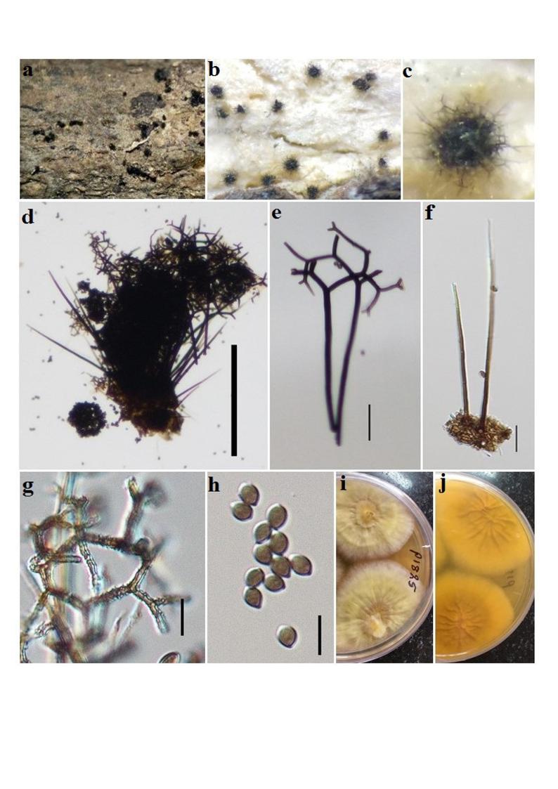
Fig. Dichotomopilus ramosissimus (AMH-10059, new host record). a–c Ascomata on host. d, e Mature ascomata on lacatophenol mount. f, g Terminal ascomatal hairs. h Ascospores. Scale bars: d = 200 μm, e = 100 μm, e, f = 20 μm, g, h = 10 μm.