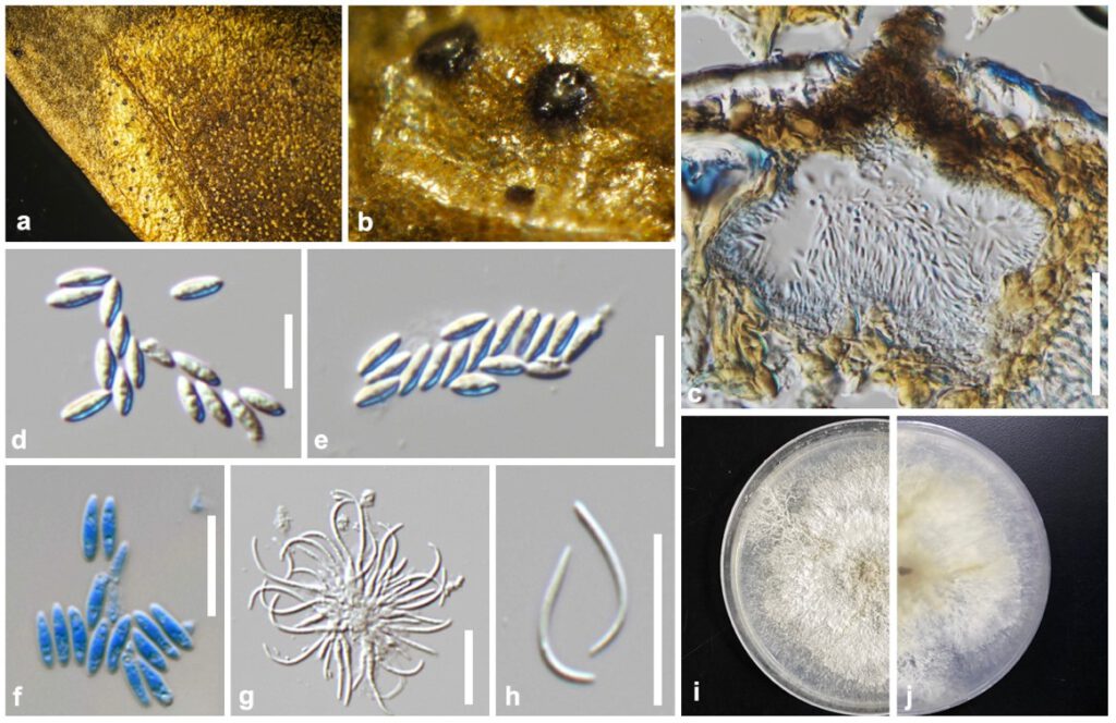
Diaporthe viridula Y.Y. Chen, A.J. Dissanayake and Jian K. Liu sp. nov. (Figure d)
MycoBank number: MB; Index Fungorum number: IF; Facesoffungi number: FoF 12615;
Etymology: Refers to the pigment color in peridium.
Holotype: GZAAS 22-0034
Saprobic on decaying leaf. Sexual morph: Not observed. Asexual morph: Conidiomata 200–300 μm diam., globose to irregular, erumpent at maturity. Peridium up to 30–80 μm diam., parenchymatous, consisting of 3–4 layers of medium brown textura angularis. Conidiophores 10–20 × 2–2.4 μm, cylindrical, hyaline, smooth, branched, ampulliform, straight to sinuous. Conidiogenous cells up to 0.5–1 μm, phialidic, cylindrical, terminal, with slight tapering towards apex. Paraphyses 10–20 × 1–2 μm, abundant among conidiophores. Alpha conidia (5–)7–8(–9) × 2–3 μm ( = 7 × 3, n = 30), aseptate, hyaline, smooth, ovate to ellipsoidal, biguttulate, base subtruncate. Beta conidia (20–)24–28(–30) × (3–)3.5–4(–4) μm ( = 25 × 3, n = 30), aseptate, hyaline, smooth, fusiform or hooked, base subtruncate.
Culture characteristics: Colonies covering entire PDA Petri dishes after 10 d at 25 °C producing dirty white with patches of olivaceous grey and isabelline. Reverse with patches of dirty white, brown vinaceous mycelium.
Material examined: China, Guizhou Province, Xingyi city, saprobic on decaying leaf, June 2020, Y. Y. Chen (GZAAS 22-0034, holotype); ex-type living culture CGMCC = GZCC 22-0033; ibid, DuShan county, saprobic on decaying woody host, May 2020, Y. Y. Chen (GZAAS 22-0051, paratype), living culture GZCC 22-0050.
Notes: The phylogenetic results showed that two isolates of Diaporthe viridula clustered closer to D. arecae and D. eugeniae forming a distinct lineage (Figure 2) with maximum support (ML/MP/BI = 100/100/1.0). Diaporthe viridula can be distinguished from D. arecae in the concatenated alignment by 32/512 in ITS, 2/413 in tef, 14/453 in tub, 19/506 in cal and 10/515 in his while from D. eugeniae by 17/512 in ITS, 5/413 in tef, 13/453 in tub, 12/506 in cal and 12/515 in his. Though D. arecae and D. eugeniae were introduced (both are comb. nov species) by Gomes et al. (2013) they have not provided the morphological details.

Figure d. Diaporthe viridula (GZAAS 22-0034, holotype). a, b Conidiomata on host surface. c Section of conidiomata. d, e Alpha conidia. f Alpha conidia stained in methylene blue. g Beta conidia attached to conidiophores. h Beta conidia. i 10 days old culture on PDA from above. j 10 days old culture on PDA from reverse. Scale bars: c = 100 μm; d–f = 10 μm; g, h = 20 μm.
