Diaporthe lonicerae Dissanayake, Camporesi & K.D. Hyde, sp. nov., Index fungorum number: IF553189
Etymology: The specific epithet lonicerae is based on the host genus (Lonicera).
Saprobic on dead aerial branch of Lonicera sp. Sexual morph: Not observed. Asexual morph: Conidiomata up to 680 μm in diameter, superficial, solitary, scattered on PDA, globose, dark brown to black, clustered in groups of 2–5 pycnidia. Peridium 15–60 μm thick, inner layer composed of light brown textura angularis, outer layer composed of dark brown textura angularis. Conidiophores 21–35 × 1.5–2.5 μm (x̅ = 27 × 2 μm), cylindrical, aseptate, densely aggregated, straight or sinuous, terminal, slightly tapered towards the apex. Conidiogenous cells 8–11 × 2–3 μm hyaline, subcylindrical, straight to curved, tapering towards the apex. Alpha conidia 12.5–16 × 3.5–4 μm (x̅ = 14.5 × 4 μm) hyaline, biguttulate, fusiform or oval, both ends obtuse. Beta conidia 32–39 × 1–1.5 μm (x̅ = 36 × 1.5 μm) hyaline, aseptate, filiform, hamate, tapering towards both ends.
Culture characteristics: Colonies on PDA covering entire Petri dishes after 10 days, flat, with an entire edge, aerial mycelium forming irregular concentric rings with cottony texture, olivaceous-buff, isabelline to honey on surface.
Material examined: ITALY, Forlì-Cesena Province, Predappio Alta, on dead aerial branch of Lonicera sp. (Caprifoliaceae), 28 Febrary 2015, Erio Camporesi; (MFLU 15-3511, holotype); ex-type living culture MFLUCC 17-0963.
Notes: Diaporthe lonicerae clusters closer to D. saccarata, D. canthi and D. hickoriae. Phylogenetically, D. saccarata is the closest species to D. lonicerae, differing by 107 nucleotides in the concatenated alignment, in which 19 were distinct in the ITS region, 34 in the TEF region, 16 in the BT region and 38 in the CAL region. Both species possess beta conidia and morphologically, D. saccarata differs from D. lonicerae, in having 1-septate alpha conidia (Mostert
et al. 2001).
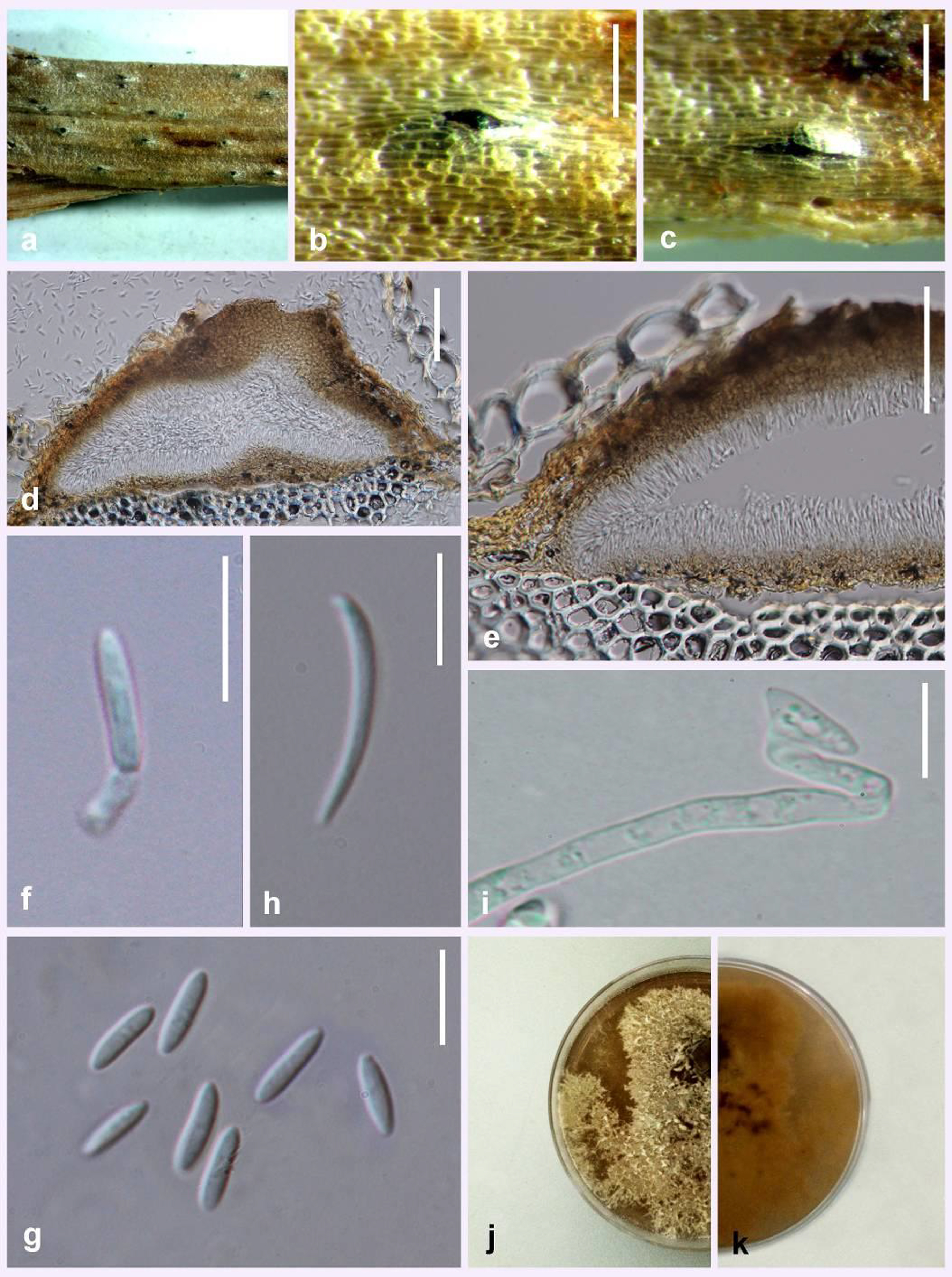
Fig. Diaporthe lonicerae (MFLU 15-3511, holotype). a–c Conidiomata on host surface. d Cross section of conidioma. e Peridium. f Alpha conidium attached to conidiogenous cells. g Alpha conidia. h Beta conidium. i Germinating conidium. j, k Culture on PDA after two weeks. Scale bars: b, c = 1 mm, d, e = 100 μm, f, g = 15 μm.