Diaporthe garethjonesii Dissanayake, Tangthirasunun & K.D. Hyde, sp. nov., Index Fungorum number: IF551403
Etymology: In reference to the significant contribution of E.B. Professor Gareth Jones made to Thai mycology.
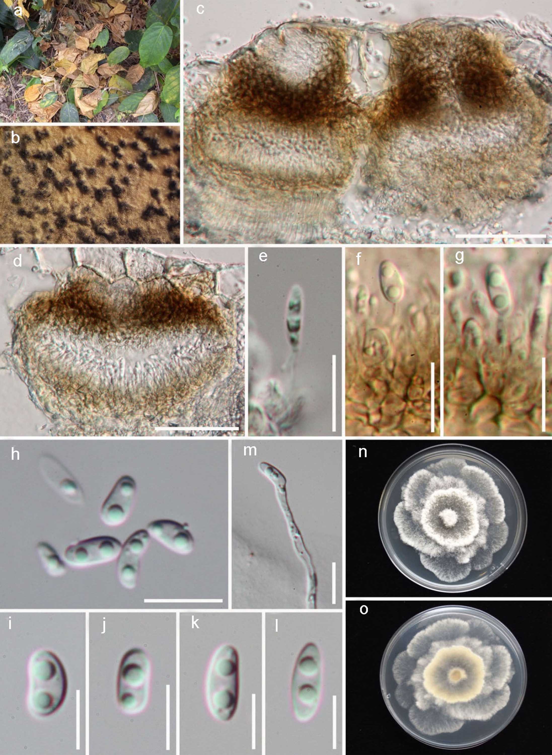
Saprobic on dead leaves. Sexual morph Undetermined. Asexual morph Conidiomata 85–125 lm diam. x 80–100 lm high (x = 115 x 85 μm, n = 10), associated with necrotic leaf tissue; pycnidial, globose, unilocular, black, immersed to sub-immersed, ostiolate, walls consisting of 5–6 layers of dark brown cells of textura angularis. Conidiophores 5–12 x 1–1.5 μm, hyaline, smooth, cylindrical, straight to sinuous. Conidiogenous cells 0.5–1 μm diam., phialidic, cylindrical, terminal and lateral, with slight taper towards apex. Paraphyses not observed. Alpha conidia 5–6 x 2–3 lm, aseptate, hyaline, smooth, guttulate, fusoid to ellipsoid, tapering towards both ends, straight, apex subobtuse. Beta conidia 40–50 x 3–4 μm hyaline, smooth, less common than alpha conidia, straight, curved or hamate.
Culture characteristics: Colonies with sparse aerial mycelium covering the dish after 2 weeks in the dark at 28 ºC. On PDA buff, honey to isabelline, reverse smokegrey.
Material examined: THAILAND, Kanjanaburi, on dead leaf, 5 May 2012, Jayarama Bhat (MFLU 13-0261, holotype), ex-type living cultures MFLUCC 12-0542a, KUMCC15-0117.
Notes: Diaporthe garethjonesii forms a well-supported clade in our phylogenetic analysis with high bootstrap and Bayesian values. The BLAST comparison of the ITS sequence of D. garethjonesii showed a 99 % match to a fungal endophyte from Hong Kong (DQ485955) (Identities = 543/548 (99 %), Gaps = 4/548 (0 %)), a 98 % match to the ITS sequence of USA isolate NY8658c (HQ108026) (Identities 537/550 (98 %), Gaps 4/550 (0 %)). The distinct clade that D. garethjonesii forms in the phylogenetic analysis, represents a separate species.
Fig. Diaporthe garethjonesii (MFLU 13-0261, holotype). a Diseased leaves. b Conidiomata on the host surface. c Longitudinal section of conidiomata. d Conidiomata on host. e–g Conidiogenous cells with developing conidia. h–m Conidiogenous cells with developing conidia stained with lactophenol cotton blue. n Alpha conidia. o Beta conidia. p Germinating conidium. q, r Colonies on PDA, q from above, r from below. Scale bars c, = 200 μm, e–p = 10 μm