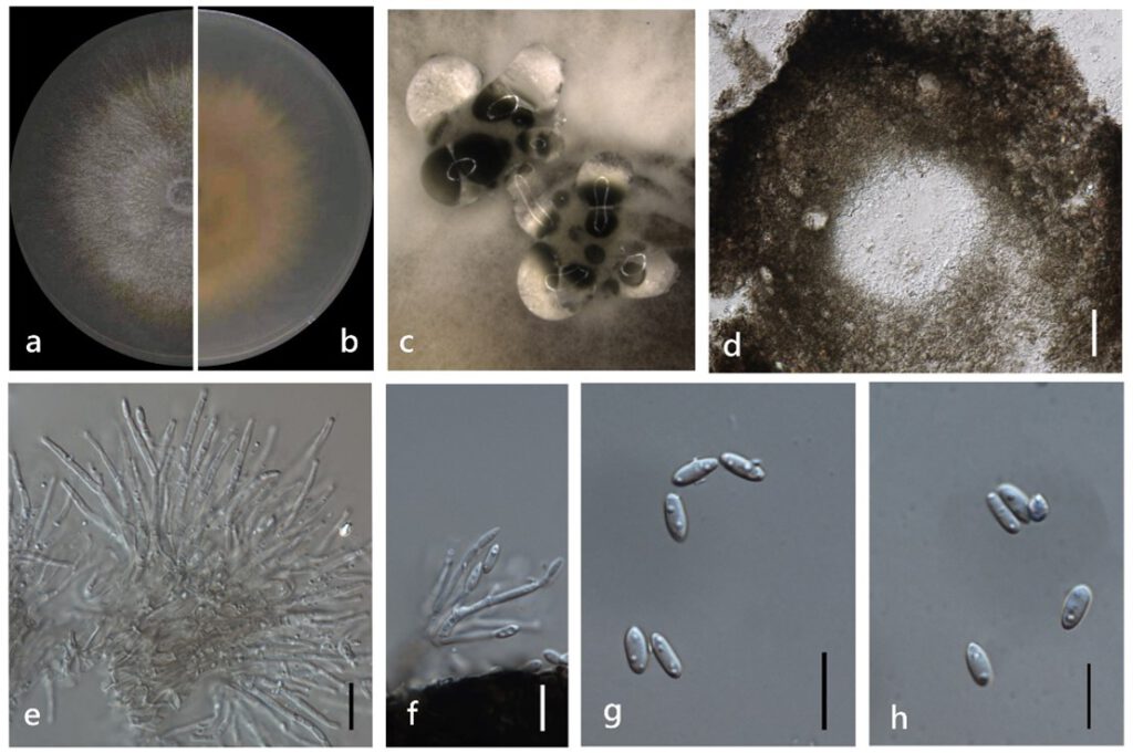
Diaporthe chongqingensis Y.S. Guo & G.P. Wang, in Guo, Crous, Bai, et al., Persoonia 45: 146 (2020) (Figure 4)
MycoBank number: MB 830656; Index Fungorum number: IF 830656; Facesoffungi number: FoF 11350;
Endophytic on Morinda officinalis stems. Sexual morph: not observed. Asexual morph: Pycnidia 130–1400 μm × 120–900 μm (`x = 542 ± 415 μm × 388 ± 267 μm) oblate or subglobose, grey to black, single or multiple cavities, translucent to black conidial drops exuded from the ostioles. Pycnidia wall thick, exuding creamy to black conidial droplets from ostioles. Conidiophore hyaline, smooth, septate, densely aggregated, cylindrical, straight to sinuous, swelling at the base, tapering towards the apex. Conidiogenous cell hyaline, cylindrical, straight, inner wall buds produce sporulation in bottle form. Alpha conidia 5–10 μm × 2–3 μm (`x = 7 ± 0.5 μm × 3 ± 0.2 μm) hyaline, aseptate, fusiform or ellipsoid, biguttulate or mlti-guttulate. Beta and gamma conidia not observed.
Culture characteristics: Colonies on PDA reach 85 mm diam. after 5 days. White cotton flocculent aerial mycelium, while the perimeter edge sparse hyphae, then ochre brown mycelium with several black conidiomata for 15 days and a lot of darker black conidiomata at 30 days. Reverse white and become reddish-brown.
Material examined: China, Guangdong Province, Zhaoqing, isolated from a healthy stem of Morinda officinalis. June 2020, W. Guo (dried culture (ZHKU 22-0032) and living culture (ZHKUCC 22-0043).
Habitat and host: Pear pyrifolia [10].
Known distribution: China [10].

Figure 4. Diaporthe chongqingensis (ZHKUCC 22-0043) (a) upper view of colonies on PDA; (b) Reverse view of colonies on PDA; (c) Pycnidia with conidial droplets on PDA; (d) a vertical Ssection through a pycnidia; (e,f) Conidiogenous cells; (g,h) Alpha conidia. Scale bars: (d) =100 µm; (e–h) =10 µm.
