Diaporthe chiangmaiensis N.I. de Silva, Lumyong & K.D. Hyde, sp. nov.
MycoBank number: MB ; Index Fungorum number: IF; Facesoffungi number: FoF 10724; Fig. **
Etymology: Name reflects the location where the specimen was collected, Chiang Mai Province, Thailand.
Holotype: MFLU 18-1305
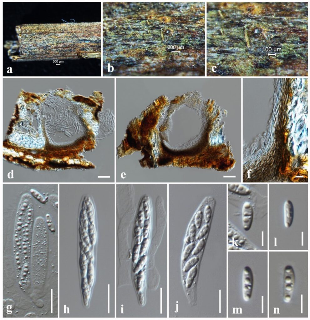
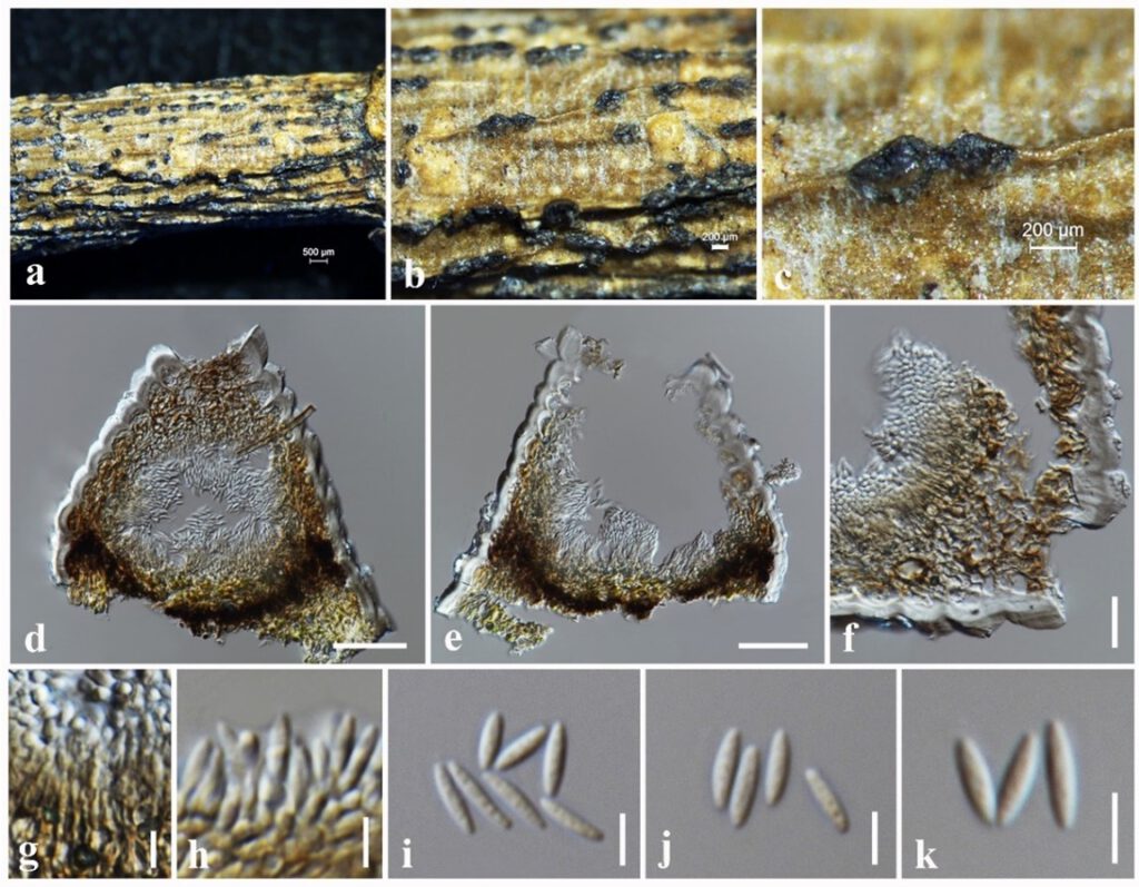
Saprobic on dead twigs attached to the host plant of Magnolia champaca. Sexual morph: Ascomata 230–270 μm high × 200–220 μm diam. ( = 240 × 210 µm, n = 10), brown, subglobose, semi-immersed, mostly immersed, solitary, scattered, coriaceous. Peridium 20–40 μm wide, composed of several layers of hyaline, brown, cells of textura angularis. Hamathecium aparaphysate or sometimes with a few cellular paraphyses. Asci 52–58 × 8–11 ( = 55 × 9 µm, n = 20), 8-spored, unitunicate, cylindrical, apex rounded with short pedicellate. Ascospores 8–10 × 2–4 ( = 9 × 3 µm, n = 30), biseriate, hyaline, fusiform to ellipsoid, 1-septate, mostly 4 guttules. Asexual morph: Coelomycetous. Conidiomata 180–200 μm high × 160–180 μm diam. ( = 194 × 172 µm, n = 10), pycnidial, dark brown, ovoid, subglobose, immersed to semi-immersed, erumpent at maturity. Conidiomata wall 35–50 μm wide, composed of 6–8 layers of pale brown cells of textura angularis. Hamathecium aparaphysate. Conidiophores 8–12 × 1–2 μm, hyaline, light brown, cylindrical, straight, smooth, densely aggregated. Conidiogenous cells 3–5 × 2–3 μm, phialidic, terminal, hyaline, cylindrical, slightly tapering towards the apex. Alpha conidia 7–9 × 1.5–3 ( = 8 × 2 µm, n = 30), hyaline, fusiform, aseptate, smooth, tapering towards both ends, straight to slightly curved.
Culture characteristics – Colonies on PDA reaching 30 mm diameter after 2 weeks at 25°C, colonies from above: circular, margin crenate, dense, fluffy appearance, white; reverse: pale brown at the margin, dark brown in the centre.
Material examined – THAILAND, Chiang Rai Province, dead twigs attached to host plant of Alstonia scholaris (Apocynaceae), 25 April 2019, N. I. de Silva, AS19 (MFLU 21-0211), living culture, MFLUCC 21-0212, THAILAND, Chiang Mai Province, dead twigs attached to host plant of Magnolia lilifera (Magnoliaceae), 11 February 2019, N. I. de Silva, NI207 (MFLU 18-1305), living culture, MFLUCC 18-0544, healthy leaves of Magnolia lilifera (Magnoliaceae), 11 February 2019, N. I. de Silva, GMT8 (MFLU 20-0606), living culture, MFLUCC 18-0935.
Notes – Phylogenetically, two saprobic strains MFLUCC 18-0544, MFLUCC 21-0212 and an endophytic strain MFLUCC 18-0935 are monophyletic with 98% ML and 1.00 BYPP statistical support (Fig. 4). This clade has a sister relationship to Diaporthe cf. heveae 2 (CBS 681.84) with 77% ML and 1.00 BYPP statistical support (Fig. 4). Diaporthe cf. heveae 2 (CBS 681.84) were isolated from leaves on Hevea brasiliensis in India (Gomes et al. 2013). Diaporthe cf. heveae 2 (CBS 681.84) was a sterile strain, and thus its morphology was not described. It is revealed that five base pair differences in ITS (500 bp) and three base pair differences in tef1 (300 bp) gene regions within our three new strains. Therefore, we recognize these three new strains belong to one species, which we introduce Diaporthe chiangmaiensis as a new species herein. We established sexual-asexual connection of D. chiangmaiensis in the current investigation of saprobic fungi recovered from dead twigs of Magnolia lilifera (Magnoliaceae).

Figure 5 – Diaporthe chiangmaiensis (NI207, holotype) a The specimen. b, c Appearance of ascomata on the substrate. d, e Sections through ascoma. f Peridium. g–j Asci. k–n Ascospores. Scale bars: a = 500 μm, b = 200 μm, c = 100 μm, d, e = 50 μm, f = 10 μm, g–j = 20 μm, k–n = 5 μm.

Figure 6 – Diaporthe chiangmaiensis (AS19) a The specimen. b, c Appearance of conidiomata on the substrate. d, e Sections through conidioma. f Peridium. g Conidiogenous cells. h–j Alpha conidia. Scale bars: a = 500 μm, b, c = 200 μm, d, e = 50 μm, f = 20 μm, g–j = 5 μm.
