Diaporthe asheicola L. Lombard & Crous, Phytopath. Mediterr. 53: 93 (2014)
Index Fungorum number: IF 807598
Saprobic on Fraxinus pennsylvanica. Asexual morph: Coelomycetous. Conidiomata up to 530 μm wide and 290–420 μm tall, (x̅ = 345 μm, n = 10), pycnidial, eustromatic, subepidermal, immersed to erumpent, globose, flask-like to conical, brown to black, scattered or aggregated, unilocular, smooth at outer surface, 1-ostiolate, including necks. Pycnidial wall consisting two regions of textura angularis; the outer region 8–15 μm wide, black, 4–5 cells thick, inner region 7–12 μm wide, brown, 3–4 cells thick, with the outside cells compressed. Conidial mass white, pale-yellow to yellow, predominantly pale-yellow, globose or in cirrhi. Alpha conidiophores 8–35 × 2–3 μm (x̅ = 27 × 2 μm, n = 20), cylindrical, filiform, aseptate and rarely branched. Alpha conidiogenous cells 3–15 × 1–2 μm (x̅ = 10 × 1.5 μm, n = 20), tapering towards the apex, subcylindrical, phialidic, collarettes and periclinal thickening present. Alpha conidia 6–9 × 2–3 μm (x̅ = 8 × 2 μm, n = 20), commonly found, fusoid to ellipsoidal, obtuse to subtruncateat base, acutely rounded at apex, multi-guttulate with guttules grouped at the polar ends, rarely biguttulate. Beta conidiophores 10–27 × 1–2 μm (x̅ = 16 × 1.5 μm, n = 20), ampulliform to subcylindrical, rarely branched. Beta conidiogenous cells 7–14 × 1–2 μm (x̅ = 11–1.5 μm, n = 20), subcylindrical, tapering towards the apex, phialidic, collarette and periclinal thickening present. Beta conidia 20–25 × 1–2 μm (x̅ = 23–1 μm, n = 20), less common than alpha conidia, straight, curved or hamate. Gamma conidia not observed. Sexual morph: Undetermined.
Material examined – Russia, Rostov region, Rostov-on-Don City Botanical Garden of Southern Federal University, on stem of Fraxinus pennsylvanica (Oleaceae), 30 May 2015, T.S. Bulgakov T823 (MFLU 15-2966).
GenBank numbers – ITS: MF443248, β-TUB: MF443249, CAL: MF443250.
Known distribution (based on molecular data) – Chile (Lombard et al. 2014).
Known hosts (based on molecular data) – Vaccinium ashei (Lombard et al. 2014).
Notes – Lombard et al. (2014) introduced D. asheicola, but this species did not sporulate on any of the media used in their study, nor on sterilised Vaccinium myrtillus tissue placed on water agar. This species lacks any data related to morphological characters due to the age or repeated sub-culturing. In our study, a Russian specimen of Diaporthe revealed molecular similarity to D. asheicola in a combined gene phylogenetic analysis (Fig. 38). Since the original description of D. asheicola by Lombard et al. (2014) lacks morphological characters, we suggest this specimen be considered as the morphological description of D. asheicola. We could not obtain a culture from a single spore and DNA was extracted directly from the fruiting bodies.
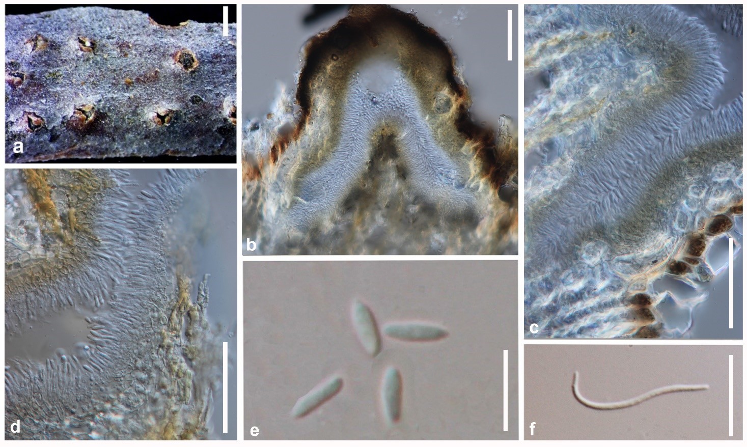
Figure 1 – Diaporthe asheicola (MFLU 15-2966, new host record). a Conidiomata on host surface. b Cross section of conidioma. c Peridium. d Alpha conidium attached to conidiogenous cells. e Alpha conidia. f Beta conidium. Scale bars: a = 0.5 mm, b–d = 200 µm, e, f = 10 µm.