Cytospora salicina Norphanphoun, Bulgakov, T.C. Wen & K.D. Hyde, sp. nov., Index Fungorum number: IF552611
Etymology: The specific epithet ‘salicina’ refers to the host plant genus Salix, on which the type specimens were collected.
Holotype: MFLU 15-2212
Associated with twigs and branches of Salix alba L. and Salix × rubens Schrank [S. alba L. × S. fragilis L.]. Sexual morph: Undetermined. Asexual morph: Conidiomata 1200–1500 × 550–600 μm diameter, semi-immersed in host tissue, solitary, scattered, erumpent, discoid, circular to ovoid, with 2–5 locules, and ostiolar neck. Ostioles 300–350 μm. at the same level as the disc surface. Peridium comprising several layers of cells of textura angularis, with inner most layer thick, dark brown, outer layer dark brown to black. Conidiophores branched or occasionally branched at the base, reduced to conidiogenous cells. Conidiogenous cells blastic, enteroblastic, phialidic, hyaline, smooth-walled. Conidia (3.6–)4.2–4.7 × 1–1.1(–1.3) μm (x̅ = 4.3 × 1 μm, n = 30), unicellular, allantoid to subcylindrical, hyaline, smooth-walled.
Culture characteristics: Colonies on MEA, reaching 9 cm diameter, after 7 days at 25 °C, producing dense mycelium, circular, margin rough, white, lacking aerial mycelium.
Material examined: RUSSIA, Rostov Region, Krasnosulinsky District, Donskoye forestry, riparian forest, on dead and dying branches of Salix alba L. (Salicaceae), 18 June 2015, T. Bulgakov, T-508 (MFLU 15-2212, holotype, KUN, isotype), ex-type living culture, MFLUCC 15-0862, KUMCC; RUSSIA, Rostov Region, Krasnosulinsky District, Donskoye forestry, ravine spinney, on dying twigs and branches of Salix × rubens Schrank [S. alba L. × S. fragilis L.] (Salicaceae), 27 October 2015, T. Bulgakov, T-1017 (MFLU 15-3679, KUN), living culture, MFLUCC 16-0637, KUMCC.
Notes: Cytospora salicina (MFLUCC 15-0862, MFLUCC 16-0637) was found on Salix spp. causing canker disease. Cytospora salicina is most similar to C. chrysosperma in having multi-locules, but differs in having smaller conidia (4.6 × 1.2 μm) and a peridium comprising several layers. Phylogenetic analysis based of combined ITS, LSU, RPB2 and ACT sequence data indicates that C. salicina can be distinguished from C. chrysosperma (HMBF17, HMBF151, HMBF158, HMBF159), C. melnikii (MFLUCC 15-0851, MFLUCC 16-0635) and C. salicacearum (MFLUCC 16-0509, MFLUCC 16-0587, MFLUCC 15-0861, MFLUCC 16-0576).
In ITS, Cytospora salicina differs from C. chrysosperma with 28 polymorphisms, three polymorphisms from C. melnikii and a single polymorphism from C. salicacearum. In RPB2 it differs in 32 polymorphisms from C. chrysosperma, 30 polymorphisms from C. melnikii and 29 polymorphisms from C. salicacearum. In ACT it differs with ten polymorphism from C. melnikii and ten polymorphisms from C. salicacearum.
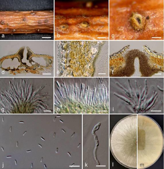
FIG: Cytospora salicina on Salix alba (MFLU 15-2212, holotype). a Stromatal habit in wood. b Fruiting bodies on substrate. c Surface of fruiting bodies. d Cross section of the stroma showing conidiomata. e Peridium. f Ostioles. g–i Conidiogenous cells with attached conidia. j Mature conidia. k Germinating spore. l, m Colonies on MEA (l-from above, m-from below). Scale bars: a = 2000 μm, b = 500 μm, c, d = 200 μm, e = 20 μm, f = 50 μm, j, k = 10 μm, g, h, i = 5 μm.