Cytospora parasitica Norphanphoun, Bulgakov, T.C. Wen & K.D. Hyde, in Ariyawansa et al., Fungal Diversity: 75(1): 146 (2015), Index Fungorum Number: 551378
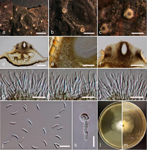
Associated with twigs and branches of Malus domestica Borkh. Sexual morph: Undetermined. Asexual morph: Conidiomata 1700–2000 × 900–1200 μm diameter, semi-immersed in host tissue, scattered, erumpent, multi-loculate, with ostiolar neck. Ostioles 320–400 μm diameter, at same level as the disc surface. Peridium comprising a few to several layers of cells of textura angularis, with most layer thin, brown to dark brown. Conidiophores unbranched or occasionally branched at the base, reduced to conidiogenous cells. Conidiogenous cells blastic, enteroblastic, phialidic, formed from the inner most layer of pycnidial wall, hyaline, smooth-walled. Conidia (5.5–)6.9–7.3 × 1.5–1.7(–2) μm (x̅ = 6.8 × 1.8 μm, n = 30), unicellular, elongate-allantoid, hyaline, smooth-walled.
Culture characteristics: Colonies on MEA, reaching 4 cm diameter, after 7 days at 25 °C, producing dense mycelium, circular, margin rough, white, lacking aerial mycelium.
Material examined: RUSSIA, Rostov Region, Shakhty City, Grushevka steppe slopes near Grushevsky pond, ravine shrubbery, on dying branches (necrotrophic) of Malus domestica (Rosaceae), 14 May 2015, T. Bulgakov (MFLU 15-1991, PDD), living culture, MFLUCC 16-0507, KUMCC.
Notes: Cytospora parasitica Norphanphoun, Bulgakov & K.D. Hyde was introduced by Ariyawansa et al. (2015) from Malus domestica in Russia (Ariyawansa et al. 2015). The morphology of this collection (MFLUCC 16-0507) is similar to C. parasitica (MFLUCC 14-1055) in having multi-loculate conidiomata and allantoid to slightly curved, unicellular, hyaline, 7 × 1.75 μm conidia (Ariyawansa et al. 2015). In phylogenetic analyses, C. parasitica (MFLUCC 16-0507) groups with the ex-type strain (MFLUCC 14-1055) of C. parasitica.
FIG: Cytospora parasitica on Malus domestica Borkh (MFLU 15-1991). a Stromatal habit in wood. b Fruiting bodies on substrate. c Surface of fruiting bodies. d Cross section of the stroma showing conidiomata. e Peridium. f Ostiolar neck. g–i Conidiogenous cells with attached conidia. j Mature conidia. k Germinating spore. l, m Colonies on MEA (l-from above, m-from below). Scale bars: a = 2000 μm, b = 1000 μm, c= 500 μm, d = 300 μm, e = 30 μm, f = 200 μm, g, h, i, j, k = 10 μm.