Cytospora cotini Norphanphoun, Bulgakov & K.D. Hyde, sp. nov., Index Fungorum number: IF552231
Etymology: from generic name of host plant, Cotinus coggygria.
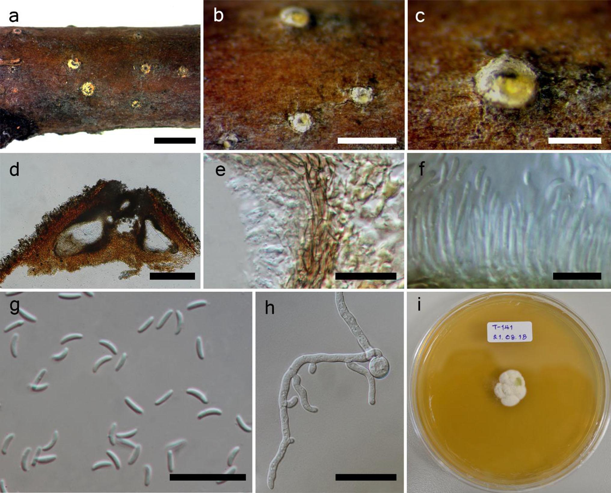
Necrotrophic on dying branches of Cotinus coggygria Scop. Sexual morph Undetermined. Asexual morph Conidiomata 800–1000 μm diam. pycnidial, solitary, immersed in host tissue, multi-locule, dark brown, ostiolate. Ostiole 250–350 μm diam. at the same level as the disc surface. Peridium comprising a few to several layers of cells of textura angularis, with inner most layer thin, hyaline, outer layer brown to dark brown. Conidiophores reduced to conidiogenous cells. Conidiogenous cells enteroblastic, phialidic, formed from the innermost layer of pycnidial wall, hyaline, smooth. Conidia (4.9–)5.6–6.5 × 0.8–1.4(–1.7) μm (x = 5.9 × 1.2 μm, n = 30), unicellular, allantoid to subcylindrical, hyaline, smooth-walled.
Culture characteristics: Colonies on MEA, reaching 1.7 cm diam. after 7 days at 25 °C, producing dense mycelium, lobate circular, with white rough margin, after 5 days, flat or effuse on the surface, without aerial mycelium.
Material examined: RUSSIA, Rostov region, Shakhty city, near Grushevsky pond, shelterbelt artificial forest, on dead branches of Cotinus coggygria Scop. (Anacardiaceae), 18 May 2014, Timur S. Bulgakov (MFLU 14-0783, holotype; KUM, isotype); ex-type living cultures, MFLUCC 14-1050, KUMCC.
Notes: Cytospora cotini is a weak pathogen on Cotinus coggygria Scop., and is often associated with Pseudocamarosporium cotinae Norphanphoun et al. The present study using morphology and phylogenetic analyses, places Cytospora cotini in Valsaceae. The new species has immersed, multi-locular conidiomata, with a single ostiole and shares common walls with the host tissue. Phylogenetic analyses, using ITS sequence data (Fig. 112), indicate that C. cotini can be distinguished from other species within the genus Cytospora. The analyses based on combined ITS, LSU, RPB2 and ACT sequence data also demonstrate that C. cotini separates from other sequenced species, and is close to Cytospora tanaitica Norphanphoun et al. However, C. tanaitica differs in having a single locule with smaller conidia than our species.
Fig. Cytospora cotini (holotype). a Appearance of fruiting bodies in wood. b Fruiting bodies on substrate. c Close up of fruiting body. d Cross section of the conidioma. e Peridium. f Conidiophores with conidia. g Conidia. h Germinating spore. i Culture characters on MEA. Scale bars a = 2 mm, b = 1 mm, c = 400 μm, d = 300 μm,
e = 20 μm, f, g = 10 μm, h = 40 μm.