Cytospora centrivillosa Senan., Camporesi & K.D. Hyde, sp., nov. MycoBank: MB821567
Etymology: Name based on two Latin words “centrum” and “villos” meaning hamathecium comprising filiform paraphyses.
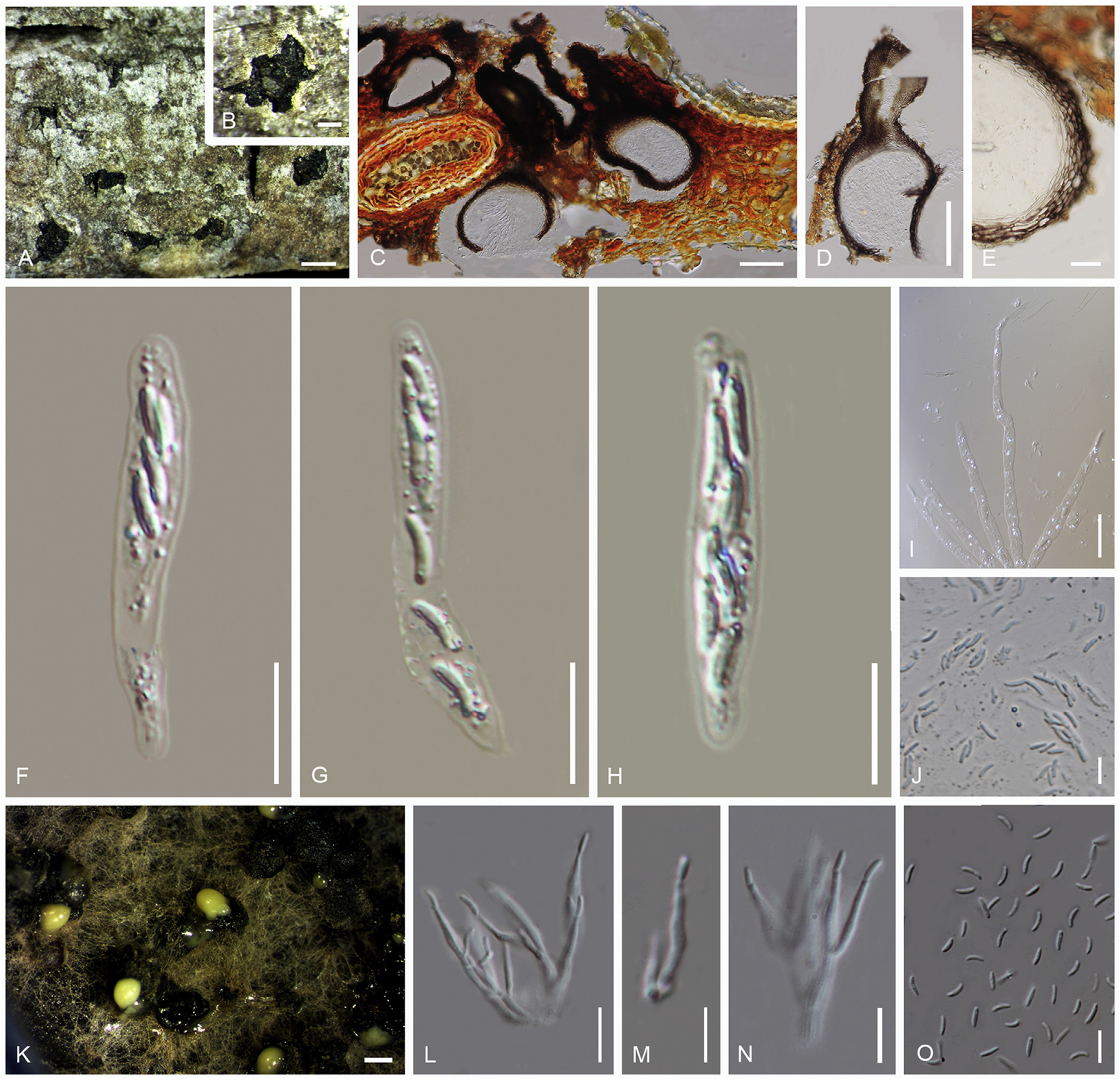
Saprobic on dead branch of Sorbus domestica. Sexual morph: Stromata poorly developed, comprising loosely packed parenchymatous cells, black. Ascomata 550–725 μm high, 160–215 μm diam (x̅ = 611 × 190 μm, n = 20), aggregated, immersed, globose to subglobose, dark brown, coriaceous, ostiolate, papillate. Papilla 285–430 μm high, 90–130 μm diam. (x̅ = 340 × 101 μm, n = 20), long, central or asymmetrically located, wall thick, internally covered by hyaline periphyses. Peridium comprises brown, thick-walled cells of textura angularis. Asci 75–85 × 15–19 μm(x̅ = 79 × 18 μm, n = 20), 8-spored, unitunicate, clavate to fusiform, without apical ring and pedicel. Ascospores 16–20 × 4–6 μm (x̅ = 17 × 5 μm, n = 20), biseriate, allantoid, hyaline, smooth. Asexual morph: Coelomycetous. Conidiomata on MEA appears as pale yellow, slimy heads of conidial mass, immersed, black. Conidiophores 6.5–8 × 3–3.5 μm (x̅ = 7.4 × 3.1 μm, n = 20), cylindrical, unbranched, hyaline. Conidiogenous cells 10–13.5 × 1–2 μm (x̅ = 11.7 × 1.6 μm, n = 20), cylindrical, tapering towards the apices, bearing single conidia at each tip, hyaline. Conidia 4–6 × 1–1.5 μm (x̅ = 5.1 × 1.1 μm, n = 20), eguttulate, allantoid, aseptate, hyaline.
Culture characteristics: Colonies growing on MEA attenuated 1 cm incubated at 18 °C within 4 d, fast growing, circular, flat, entire, white, thin, tightly attached to the media, mycelia clots arrange radially from centre to margin.
Specimens examined: Italy, Province of Forlì-Cesena, Predappio, Monte Mirabello, on dead and aerial branch of Sorbus domestica (Rosaceae), 1 Oct. 2014, E. Camporesi, IT 2132 (holotype MFLU 17–0887, isotype BBH 42449, culture ex-type MFLUCC 16–1206); Province of Forlì-Cesena, Predappio, Monte Mirabello, on dead and aerial branch of Sorbus domestica (Rosaceae), 13 Oct. 2014, E. Camporesi, IT 2132B, MFLU 17–0999, culture MFLUCC 17–1660.
Note: Cytospora centrivillosa is morphologically and phylogenetically distinct from other species in Cytospora and our analysis results in a distinct clade with full support (Clade 16).
Fig. Cytospora centrivillosa (MFLU 17–0887). A, B. Stromata on substrate. C, D. Vertical cross section of ascomata. E. Peridium. F–H. Asci. I. Paraphyses. J. Ascospores. K. Conidiomata on MEA. L–N. Conidiogenous cells, conidiophores, conidia. O. Conidia. Scale bars: A = 500 mm, B = 200 mm, C, D = 50 mm, E = 5 mm, I = 20 mm, F–H, J, L–O = 10 mm, K = 500 mm.